


Sarbecovirus Evolution Tracking: Recombinant hACE2 Protein as a Key Research Tool
- 2026-03-18
- 145
Coronaviruses (CoVs) are a group of enveloped, single-stranded, positive-sense RNA viruses. Within this group, the Sarbecovirus subgenus of betacoronaviruses includes pathogenic viruses such as SARS-CoV and SARS-CoV-2, along with numerous bat-derived SARS-like coronaviruses (SL-CoVs). The ability of these viruses to infect a host hinges on whether the receptor-binding domain (RBD) of their spike (S) protein can recognize the host cell surface receptor angiotensin-converting enzyme 2 (ACE2).
However, within Sarbecoviruses, only some members can utilize ACE2 as an entry receptor, while others cannot. This raises a fundamental scientific question: How did the ability to bind ACE2 originate within this viral group? The scientific community previously debated two competing hypotheses: one suggested ACE2 binding was an ancestral trait subsequently lost in some viruses due to deletions or mutations, while the other proposed this ability could be acquired through recombination with other viruses.
To address this, a research team led by Dr. Shunyi Zhu at the Institute of Zoology, Chinese Academy of Sciences, employed a multi-faceted approach. Their work integrated phylogenetic analysis, ancestral sequence reconstruction, structural biology, and molecular dynamics simulations. This comprehensive study elucidated a novel evolutionary mechanism: the ACE2-utilizing capability in Sarbecoviruses originated through three independent and parallel evolutionary origination events. This finding provides crucial insights into the early evolution and cross-species transmission of these viruses.
I. The Structural Basis of SARS-CoV-2 RBD Binding to hACE2
Studies reveal that the receptor-binding motif (RBM) of the SARS-CoV-2 RBD is composed of three loops: RBML1, RBML2, and RBML3. Among them, RBML2 is the longest and contains an additional disulfide bond (SS4). Together, these three loops constitute the functional unit responsible for binding to hACE2. The RBM forms a stable binding interface with the α1 and α2 helices of hACE2 through extensive hydrophobic interactions and hydrogen bonds. This structural foundation serves as a critical prerequisite for Sarbecovirus entry into host cells and provides a structural reference for subsequent research investigating the impact of RBD mutations on binding affinity.
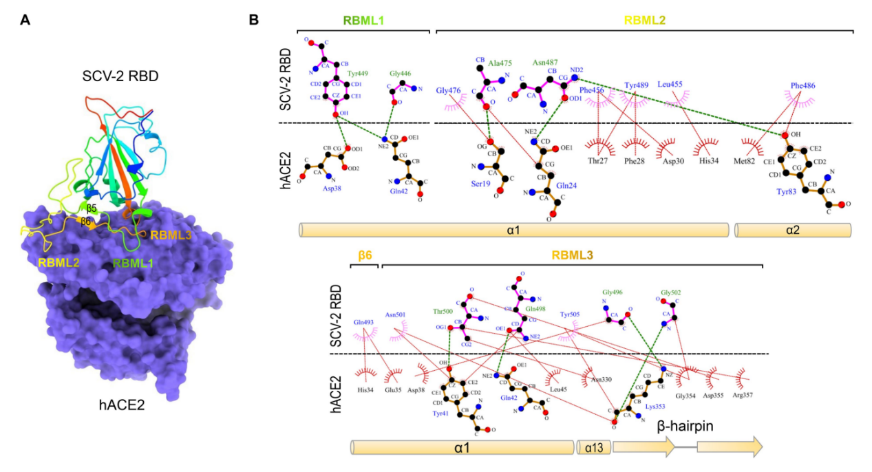
Fig 1. SARS-CoV-2 RBD binds to hACE2 via residues located on three loops
II. Design and Construction of SARS-CoV-2 RBD Mutants for Molecular Dynamics Simulation
Molecular dynamics simulations revealed that designed modifications—including loop truncation or extension, disruption of disulfide bonds, and substitution of the RBD core sequence—all impact RBD conformation and function. To ensure structural fidelity between the mutants and the native conformation, mutant models were generated via homology modeling based on the SARS-CoV-2 RBD crystal structure. This approach provided standardized subjects for subsequent precise analysis.
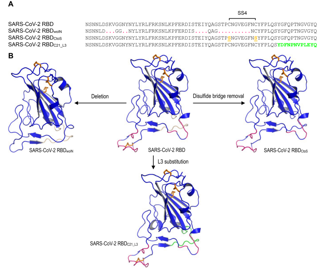
Fig 2.SARS-CoV-2 RBD mutants for MD simulations
III. Long-Loop RBD Variants Generally Possess ACE2 Binding Capacity
Phylogenetic analysis indicates that within Sarbecoviruses, RBD variants characterized by long loops typically exhibit ACE2 binding activity, whereas short-loop variants completely lack this function. Seven key divergent sites within the sequence directly determine this functional differentiation of the RBD. The mutation rate within specific loop regions of long-loop RBDs is significantly higher than in their flanking regions, demonstrating that accelerated mutation in the long-loop region is a core driver for the evolution of ACE2-binding capability.
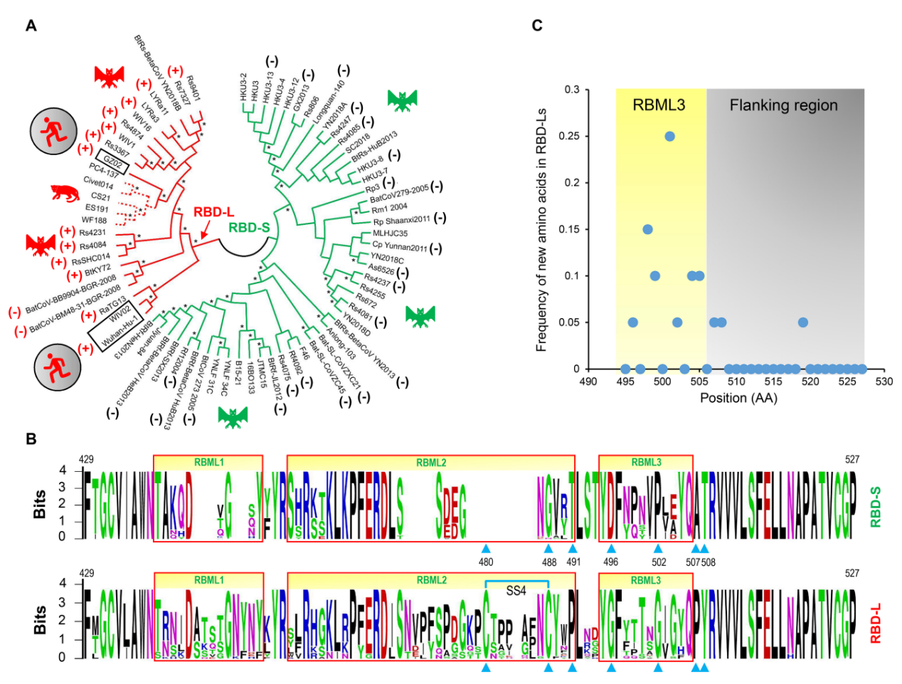
Fig 3. Classification and evolutionary conservation of Sarbecovirus RBDs
IV. Regulatory Mechanisms of Mutations on RBD Structure and Function
Dynamic simulations and conformational analyses confirm that insertion mutations establish a structural and functional adaptation mechanism, forming the conformational foundation required for ACE2 binding. This leads to a decrease in the overall structural stability of the RBD. For instance, the substitution of key residues (e.g., D496G, P502G) eliminates electrostatic repulsion and steric hindrance at the RBD-ACE2 interface while introducing new hydrogen bonds. The disulfide bond SS4 maintains effective interaction with ACE2 by switching between open and closed states and collaborates with Pro-491 to regulate the conformational plasticity of RBML2, perfectly illustrating the cooperative mechanism between insertions and point mutations.
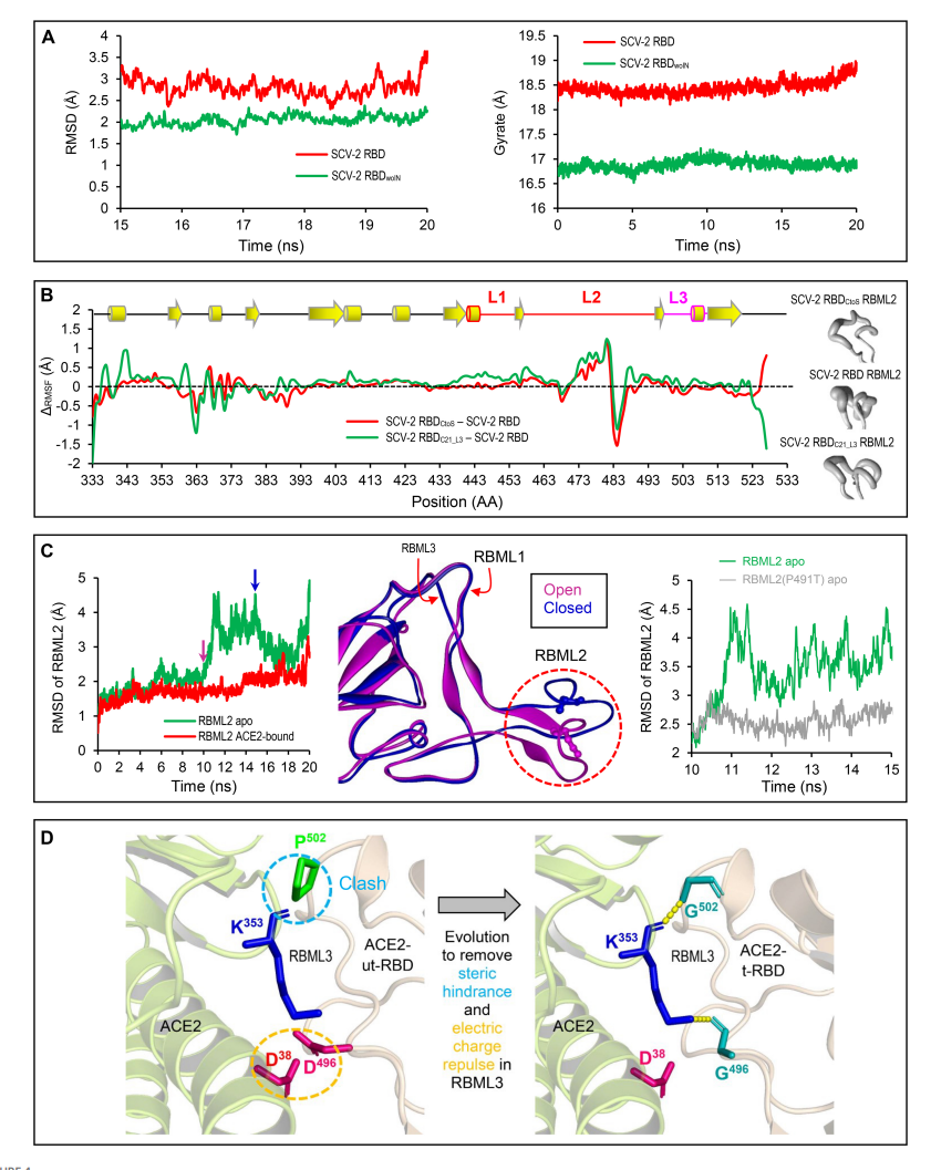
Fig 4. Structural and Dynamic Evidence for the Origin of ACE2 Binding
V. Phylogenetic Evidence for Parallel Evolution in Sarbecoviruses
Whole-genome phylogenetic analysis reveals that Sarbecoviruses are divided into three independent evolutionary lineages: Euro-African SARS-like coronaviruses, Asian novel coronaviruses, and Asian Severe Acute Respiratory Syndrome coronaviruses. Each lineage contains a mixture of virus strains carrying both long and short receptor-binding domains. Related studies suggest that attributing the origin of ACE2-binding capability to three independent insertion events provides a more plausible evolutionary explanation than attributing it to five gene deletion events.
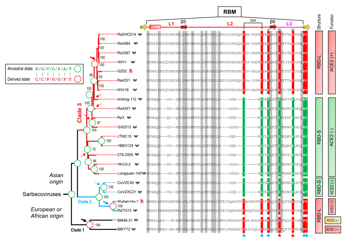
Fig 5. Evidence for the Parallel Evolution of ACE2-Binding Capacity in Three Sarbecovirus Lineages
VI. Validation of the Parallel Evolution Hypothesis through Ancestral Sequence Reconstruction
Using ancestral sequence reconstruction, FastML analysis was performed on RBM sequences lacking recombination breakpoints. The results indicate that, regardless of whether phylogenetic trees constructed from genomes containing or lacking the RBD-coding region, or different protein substitution models were used, the ancestral state of the Sarbecovirus RBD was non-ACE2-binding (RBD-S-like). Furthermore, subtrees constructed after removing the RBD-coding region retained the characteristic pattern of parallel substitutions. This excludes interference from recombination in the evolutionary analysis and further consolidates the reliability of the parallel evolution hypothesis.
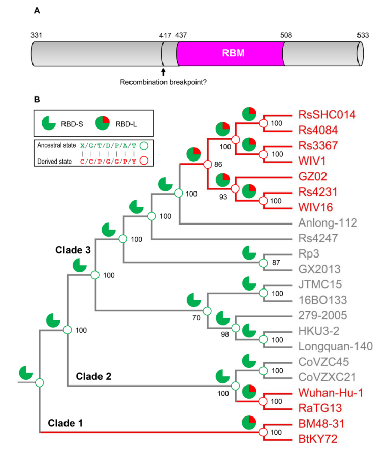
Fig 6. Ancestral sequence reconstruction elucidating the history of insertions and point mutations as proposed by the parallel evolution hypothesis
VII. Purification, Characterization, and SPR Functional Validation of Recombinant RBD
Size exclusion chromatography and SDS-PAGE results demonstrate the successful high-purity preparation of recombinant RBD and its mutants, with the actual molecular weights of the proteins matching their theoretical values. SPR analysis revealed no detectable binding response for specific bat-derived RBDs and their mutants. In contrast, SARS-CoV-2 RBD exhibited high-affinity binding to hACE2. These findings provide key functional evidence supporting the "evolutionary intermediate" hypothesis.
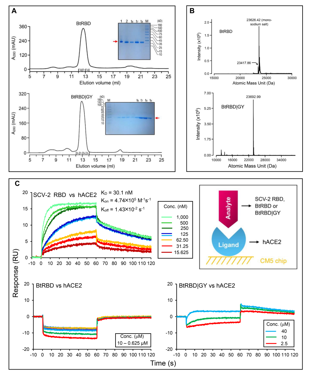
Fig 7. Purification, characterization, and functional validation of recombinant bovine serum albumin
This study confirms that ACE2-utilizing Sarbecoviruses originated through three independent, parallel evolutionary events driven by mutations. In the ancestral RBDs of the Asian SARS-CoV, Asian SARS-CoV-2, and Euro-African SL-CoV lineages, insertion mutations lengthened RBML1 and RBML2. These insertions, coupled with specific point mutations, eliminated charge repulsion and steric hindrance, thereby conferring ACE2-binding capability through transitional evolutionary intermediates. This finding elucidates the molecular pathway of viral functional innovation and provides key evidence to resolve the long-standing debate on the origin of ACE2-binding ability.
The recombinant human hACE2 protein used in this study was sourced from KMD Bioscience (Tianjin, China). The protein was prepared following professional expression protocols, resulting in a spatial conformation and post-translational modification profile highly consistent with native human protein. It serves as a reliable experimental material for research related to Sarbecovirus evolution.
KMD Bioscience is dedicated to protein expression research. Leveraging mature prokaryotic, mammalian, insect, and yeast expression systems, we offer a variety of high-purity, high-bioactivity protein products. Our alpaca nanobody platform can produce premium antibodies with pM-level affinity. We also provide complete technical solutions, from protein expression to scaled-up production processes, to accelerate the translation of your research discoveries.
[1] Gao B, Zhu S. Mutation-driven parallel evolution in emergence of ACE2-utilizing sarbecoviruses. Front Microbiol. 2023;14:1118025.
- Recombinant hACE2 Protein
- Protein Expression Services
- Recombinant Protein
- Recombinant Protein Preparation

- Protein Platform
- Protein Expression Services
- Protein Modification & Detection Services
- Protein Interaction Services
- Protein Purification Service
- Antibody Discovery Platform
- VHH Antibody Platform
- Phage Display Platform
- Antibody Humanization Platform
- Single B Cell Screening Platform
- Antibody Expression & Validation Platform
- Peptide Library Platform
- Custom Antibody Platform
- Innovative Drug Discovery Platform
- Product Center
- Proteins
- Antibodies
- Raw Materials Reagents
- Small Molecule Reagents
- Peptide Products
- Nanoparticle Products
- Contact Us
- Phone:400-621-6806
- Mobile:13110936064(WeC
- E-mail:[email protected]
- Address:Room 108, Building 10, No. 6 Hechang Road, Wuqing Development Zone, Tianjin City
-

WeChat
-

Service Account
-
 400-621-6806
 -
 3599163699
 -
 Monday to Friday 9:00-18:00
 -

-
 0
0
-
