


Introduction to Antibodies
- 2026-07-10
- 300
The currently discovered antibodies, also known as immunoglobulins, all exhibit a "Y"-shaped structure. Conventional immunoglobulins have a symmetric structure composed of four polypeptide chains: two heavy chains (H chains) and two light chains (L chains). These chains are connected by disulfide bonds and non-covalent bonds, forming a monomeric molecule consisting of four polypeptide chains. Light chains come in two types, κ and λ, while heavy chains are classified into five types: μ, δ, γ, ε, and α.Non-conventional immunoglobulins refer to camelid antibodies and cartilaginous fish antibodies. Both types lack light chains and consist solely of two heavy chains, hence they are referred to as "heavy-chain antibodies." Compared to camelid antibodies, cartilaginous fish antibodies, such as those in sharks, possess three additional pairs of heavy-chain domains (Hc domains), as illustrated in Figure 2.
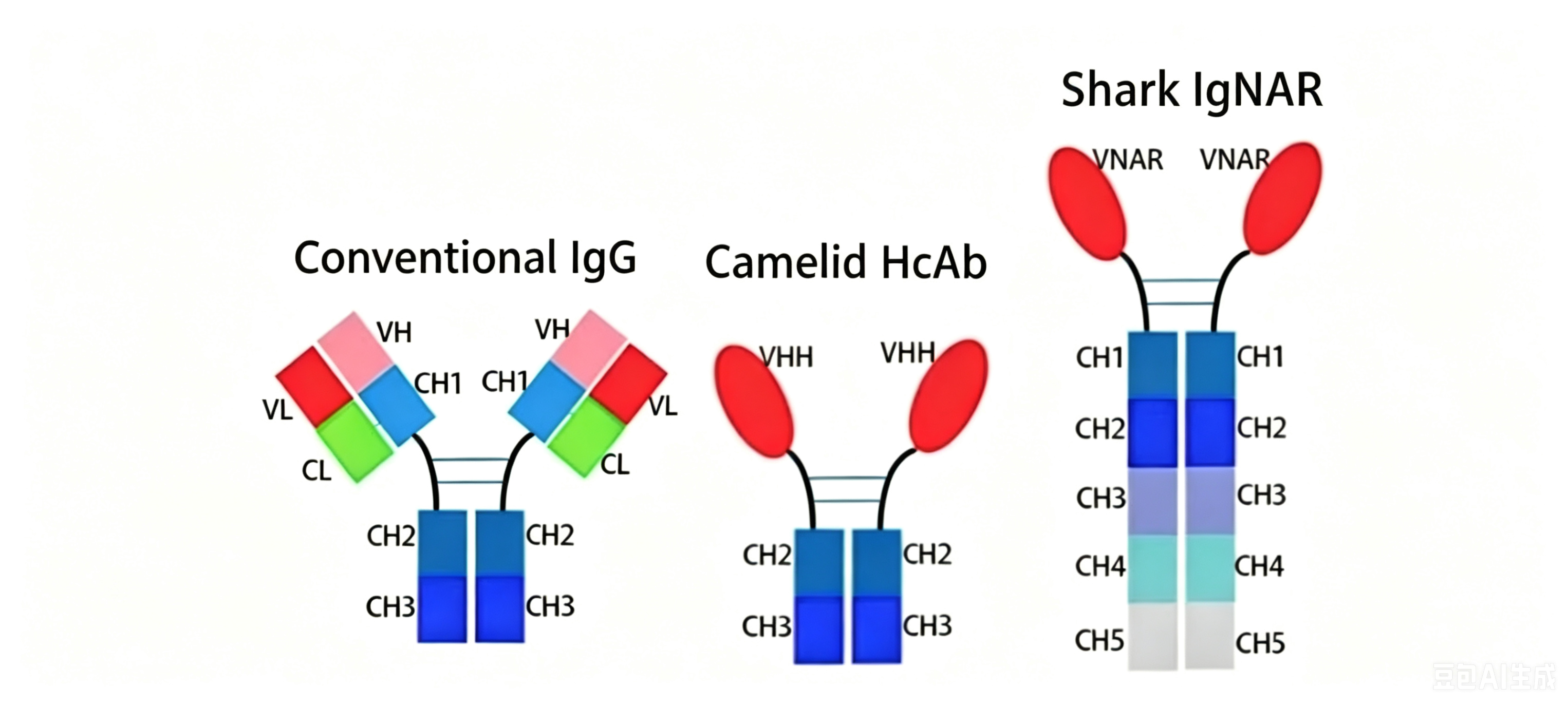
Figure 2 Conventional IgG antibody vs. heavy-chain antibodies:The conventional IgG antibody consists of a heavy chain with 8 domains and a light chain with 2 domains, pairing together to form a "Y"-shaped structure;Camelid antibodies are composed of 6 heavy-chain domains;Shark antibodies contain 12 heavy-chain domains.
1. Antibody Structure
Heavy Chain Structure: Conventional immunoglobulins have two heavy chains (H chains), each with a molecular weight of approximately 50 kDa. There are five heavy chain isotypes—μ, δ, γ, ε, and α—corresponding to the immunoglobulin classes IgM, IgG, IgA, IgD, and IgE, respectively.
Heavy-chain antibodies (HCAbs) are a unique class of antibodies that naturally lack light chains. They are found in camelids (such as the Asian Bactrian camel Camelus bactrianus, the African dromedary C. dromedarius, and South American camelids including the llama Lama glama, guanaco L. guanicoe, alpaca Vicugna pacos, and vicuña V. vicugna) and cartilaginous fishes.In 1995, a similar antigen receptor lacking light chains or associated protein molecules was discovered in cartilaginous fishes, including the nurse shark (Ginglymostoma cirratum), wobbegong (Orectolobus maculatus), and chimaera. This receptor was named the new antigen receptor (NAR) or nurse shark antigen receptor (NAR). Due to its functional similarities with immunoglobulin (Ig) subtypes in transmembrane signaling and secretion mechanisms, it is also referred to as the immunoglobulin new antigen receptor (IgNAR) (see Figure 2).The most widely utilized component of heavy-chain antibodies is the single-domain antibody (sdAb). An sdAb is a genetically engineered antibody consisting solely of the variable region (VHH) of the heavy-chain antibody. It is also known as a VHH antibody (variable domain of the heavy chain of heavy-chain antibody) or nanobody (Nb), with a molecular weight of 12–15 kDa.
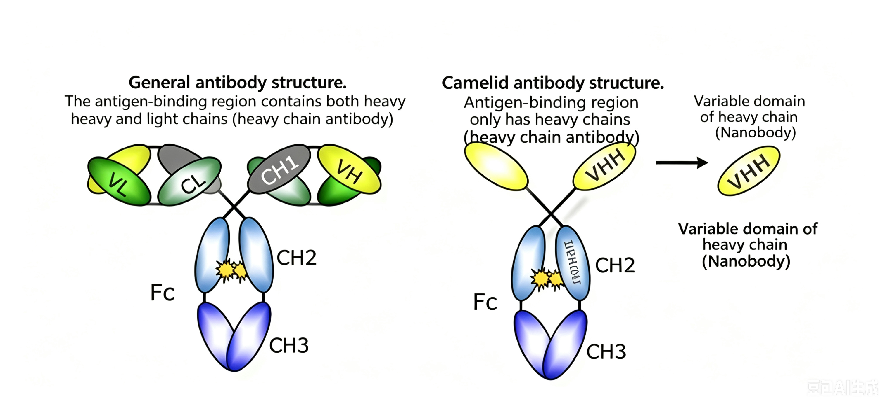
Figure 4 Schematic comparison of conventional IgG antibodies, heavy-chain antibodies, and nanobodies
2. Classification of Conventional Antibodies
Antibodies can be categorized into various types based on different classification systems. The most commonly used classification method divides conventional antibodies into five classes - IgG, IgM, IgA, IgE, and IgD - according to their physicochemical properties and biological functions.
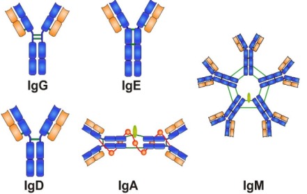
Figure 5 Classification of conventional antibodies
-- IgG is a major immunoglobulin in serum, accounting for approximately 65-75% of total immunoglobulins. It is widely distributed in tissue fluids, with roughly equal distribution between intravascular and extravascular compartments. IgG serves as an important substance for the body's anti-infection defense.
-- IgM is the first class of immunoglobulin synthesized by a mature fetus and also the earliest produced immunoglobulin following infection or immunization. Among the five immunoglobulin classes, IgM exhibits the strongest agglutination capacity, hence possessing the most potent cytotoxIC activity and cell lysing activity. Natural blood group antibodies belong to IgM, and some autoantibodies such as anti-phospholipid antibodies and rheumatoid factor (RF) are also of the IgM class. Elevated IgM antibodies in fetal umbilical cord blood indicate fetal infection.
-- IgA is relatively less abundant in serum and tissue fluids, with serum IgA comprising 15-25% of total immunoglobulins. However, it is present in higher concentrations in external secretions such as colostrum, saliva, tears, intestinal secretions, and bronchial secretions. Since IgA primarily exists in external secretions, it plays a crucial role in first-line anti-infection defense.
-- IgE has a monomeric structure and is the least abundant immunoglobulin in normal human serum. IgE is present in extremely low concentrations in serum and tissue fluids, with its main biological function being binding to specific receptors on tissue mast cells and basophils. IgE cannot activate complement. IgE levels vary significantly among normal populations but may increase in patients with specific allergic reactions or early-stage parasitic infections. When allergens react with IgE bound to these receptors, they can trigger mast cells and basophils to degranulate, releasing bioactive substances such as histamine and 5-hydroxytryptamine.
-- IgD concentration is very low in normal human serum, often barely detectable. IgD primarily exists on the surface of human B lymphocytes as antigen receptors, with serum IgD being extremely scarce and structurally different from membrane-bound IgD. The variable region of IgD on B cells is identical to that of IgG, IgA, or IgM that the cell will subsequently secrete. When antigens bind to IgD receptors, they stimulate B cells to proliferate, differentiate, and secrete other classes of antibodies specific to the antigen.
3. What are the functions of antibodies?
(1) Specific binding to antigens: Antibodies themselves cannot directly dissolve or kill target cells carrying specific antigens. They usually require the collaboration of complement or phagocytes to clear pathogenic microorganisms or cause pathological damage. However, antibodies can directly neutralize viruses by specifically binding to them or their toxins.
(2) Activation of complement: IgM, IgG1, IgG2, and IgG3 can activate the complement system via the classical pathway, while aggregated IgA, IgG4, and IgE can activate complement through the alternative pathway.
(3) Binding to cells: Different classes of immunoglobulins can bind to various types of cells, participating in immune responses.
(4) Transplacental and mucosal transfer: Immunoglobulin G (IgG) can cross the placenta and enter the fetal bloodstream, providing natural passive immunity to the fetus. Immunoglobulin A (IgA) can pass through the mucous membranes of the digestive and respiratory tracts, serving as a key factor in local mucosal immunity against infections.
- Antibody
- Antibody production
- Antibody expression
- Antibody preparation

- Protein Platform
- Protein Expression Services
- Protein Modification & Detection Services
- Protein Interaction Services
- Protein Purification Service
- Antibody Discovery Platform
- VHH Antibody Platform
- Phage Display Platform
- Antibody Humanization Platform
- Single B Cell Screening Platform
- Antibody Expression & Validation Platform
- Peptide Library Platform
- Custom Antibody Platform
- Innovative Drug Discovery Platform
- Product Center
- Proteins
- Antibodies
- Raw Materials Reagents
- Small Molecule Reagents
- Peptide Products
- Nanoparticle Products
- Contact Us
- Phone:400-621-6806
- Mobile:13110936064(WeC
- E-mail:[email protected]
- Address:Room 108, Building 10, No. 6 Hechang Road, Wuqing Development Zone, Tianjin City
-

WeChat
-

Service Account
-
 400-621-6806
 -
 3599163699
 -
 Monday to Friday 9:00-18:00
 -

-
 0
0
-
