


Production of Mouse Monoclonal Antibodies
- 2026-07-10
- 304
I. Mouse Monoclonal Antibodies
Mouse monoclonal antibodies are derived from hybridoma cells, which are generated by fusing activated B lymphocytes with myeloma cells. These hybridomas produce murine-derived monoclonal antibodies capable of both specific antibody secretion and unlimited proliferation.
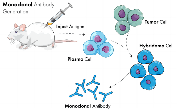
Figure 1: Monoclonal Antibody Preparation
II. Methods for Mouse Monoclonal Antibody Preparation
KMD Bioscience offers two approaches for mouse monoclonal antibody preparation services: one utilizes phage display technology to produce recombinant monoclonal antibodies, while the other employs hybridoma technology to generate hybridoma cell lines.
Traditional monoclonal antibody technology involves the fusion of myeloma cells with antibody-secreting B lymphocytes (excluding fully human monoclonal antibody preparation techniques) to form hybridoma cells that produce monoclonal antibodies. Since these antibodies target only a single epitope on the antigenic molecule, they exhibit high specificity and potency.
III. Advantages of Mouse Monoclonal Antibody Preparation
Mice are one of the most common model organisms in biology and medical research. Mouse monoclonal antibodies (mAbs) are widely used due to their high specificity and advantages such as rapid, large-scale, and continuous production.
IV. KMD Bioscience’s Diverse High-Quality Mouse Monoclonal Antibody Products
Based on a well-established antibody platform system, KMD Bioscience currently produces various types of murine monoclonal antibody products. These include tag antibodies such as HRP-labeled mouse anti-human IgG (FC) (Cat: SA2265), animal disease antibodies like mouse anti-pseudorabies virus monoclonal antibody (Cat: ANAB1024), and small molecule antibodies such as penicillin mouse monoclonal antibody (Cat: PA042). These products are widely used in various applications including WB, ELISA, and other detection assays.
V. KMD Bioscience Provides High-Quality Mouse Monoclonal Antibody Preparation Services
KMD Bioscience provides mouse monoclonal antibody preparation services, with the main process including: antigen preparation → mouse immunization → cell fusion → hybridoma screening → subcloning → ascites production → antibody purification.
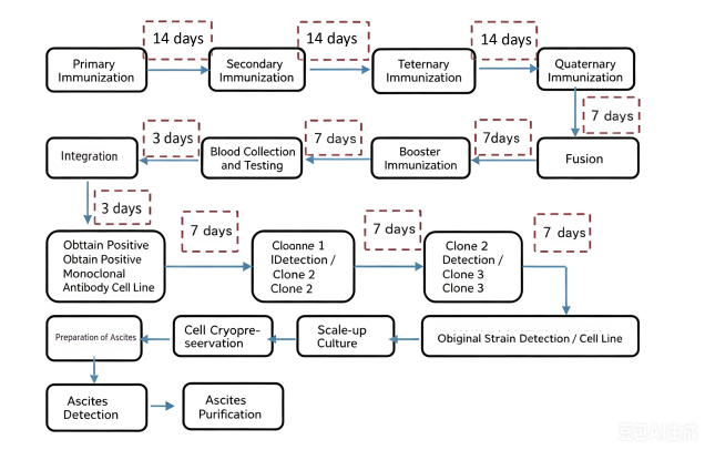
Figure 2: Mouse Monoclonal Antibody Production Process
1. Antigen Preparation
The customer provides the antigen, which can be proteins, peptides, or various small molecule antigens. These are mixed with Freund’s adjuvant for immunization.
Table 1: Mouse Monoclonal Antibody Preparation Schedule Timeline
| Process | Time | Description | Date |
| Pre-immune serum collection | Day 1 | Pre-immune serum collection: 20 μl per mouse | ****** |
| First immunization | Day 1 | 0.1 mg immunogen (CFA), subcutaneous multi-point injection (5 sites) | ****** |
| Second immunization | Day 14 | 0.05 mg immunogen (IFA), subcutaneous multi-point injection (4 sites) | ****** |
| Third immunization | Day 28 | 0.05 mg immunogen (IFA), subcutaneous multi-point injection (4 sites) | ****** |
| Fourth immunization | Day 42 | 0.05 mg immunogen (IFA), subcutaneous multi-point injection (4 sites) | ****** |
| ELISA screening | Day 49 | Whether to continue immunization is determined based on ELISA results | ****** |
| Booster immunization | Day 62 | 0.05 mg immunogen (saline), intraperitoneal injection | ****** |
| ELISA confirmation | Day 69 | Whether to continue immunization is determined based on ELISA results | ****** |
| Cell fusion & subcloning | Day 76 | If the titer is qualified: myeloma cells and splenocytes are fused, followed by clone screening | ****** |
| Stable cell line expansion | Day 194 | Clone screening | ****** |
| Ascites production & purification | Day 208 | Antibody purification by Protein A/G affinity chromatography | ****** |
2、Mouse Immunization
Prepare five Balb/c mice and administer immunogen injections at 14-day intervals. Collect blood for titer detection.
3、Cell Fusion, Screening, and Subcloning
Splenocytes from mice with serum titers meeting the requirements (protein/virus >105; peptide/small molecule >104) were fused with murine myeloma cells. The fused cells were subjected to limiting dilution and plated in 96-well plates for cloning. Hybridoma cells were selected using HAT medium, and 1–10 positive clones were identified by immunogen detection. These clones were subcloned and expanded in 96-well plates. ELISA was used to screen positive subclones, followed by further expansion.After fusion, primary cloning by limiting dilution, and supernatant screening, multiple cell lines meeting project requirements were obtained. Four cell lines were selected for subsequent ascites production.
4、Purification of Ascites Antibodies
The selected hybridoma cells were used to generate ascites, and the antibodies were purified from the ascitic fluid using Protein A/G affinity chromatography.
4.1、Treatment of Protein A/G-Sepharose Column
1) Select an appropriate chromatography column, clean it thoroughly, and fix it vertically on an iron stand.
2) Take an appropriate amount of the ethanol suspension of Protein G-Sepharose, let it stand until the resin settles, then aspirate the supernatant ethanol. Suspend the resin multiple times with binding buffer to thoroughly wash away the ethanol.
3) Mix the binding buffer with Protein A/G to form a 50-70% slurry, then pour the slurry into the column.
4) Connect a peristaltic pump and pack the column at a flow rate of 50 cm/h (equivalent to 0.7 ml/min for a column with a diameter of 1 cm). After the resin has fully settled, continue washing the column with 5-10 column volumes of binding buffer.
4.2、Sample Processing and Loading
1) Ascites sample: Centrifuge at 12,000 × *g* for 10 min, then filter the supernatant through a 0.45 μm membrane.
2) Loading: Apply the filtered sample to the affinity column at a flow rate of 0.4 mL/min.
3) After the sample has entered the resin, reduce the flow rate to 0.7 mL/min and continue washing with 10 column volumes of binding buffer until the A280 absorbance returns to baseline levels (< 0.02).
4.3、Sample Elution
1) Maintain the same flow rate and switch to elution buffer to elute the antibodies bound to the column. Collect the eluate in 2 mL fractions (2 mL/tube). Add 0.1 mL of neutralization buffer to each collection tube to promptly adjust the pH of the eluted antibody solution.
2) Measure the A280 of each fraction and stop elution when A280 falls below 0.05. Pool the peak fractions with A280 ≥ 0.2.
3) If desalting or buffer exchange is required, dialyze the sample against phosphate-buffered saline (PBS) or another specified solution.
4.4、Purity Assessment
Electrophoresis: Mix the purified protein with 5X loading buffer at a 4:1 ratio, then boil the sample at 100°C for 10 minutes. Load 10 μL onto an SDS-PAGE gel and run at 120 V for 90 minutes. Stain the gel with Coomassie Blue for 30 minutes, followed by destaining.
5、Delivery
KMD Bioscience delivers 3-5 positive hybridoma clones, along with highly specific and high-affinity purified mouse monoclonal antibodies. The delivery includes experimental reports and characterization reports featuring SDS-PAGE and ELISA test results. Additionally, based on customer requirements, we provide antibody validation data such as Western Blot, IHC, IP, IF, and other relevant assays.
Mouse monoclonal antibodies are one of the most commonly used monoclonal antibodies, with extensive applications in tumor diagnosis and therapy, cell surface antigen detection, and measurement of trace biological components. Therefore, producing highly specific, large-scale antibody preparations is a key requirement for research users.KMD Bioscience has accumulated substantial experience and technical expertise in antibody production, offering comprehensive custom mouse monoclonal antibody services covering the entire upstream and downstream workflow.
- Mouse monoclonal antibody
- mouse monoclonal antibody production
- antibody production service
- mouse monoclonal antibody production company

- Protein Platform
- Protein Expression Services
- Protein Modification & Detection Services
- Protein Interaction Services
- Protein Purification Service
- Antibody Discovery Platform
- VHH Antibody Platform
- Phage Display Platform
- Antibody Humanization Platform
- Single B Cell Screening Platform
- Antibody Expression & Validation Platform
- Peptide Library Platform
- Custom Antibody Platform
- Innovative Drug Discovery Platform
- Product Center
- Proteins
- Antibodies
- Raw Materials Reagents
- Small Molecule Reagents
- Peptide Products
- Nanoparticle Products
- Contact Us
- Phone:400-621-6806
- Mobile:13110936064(WeC
- E-mail:[email protected]
- Address:Room 108, Building 10, No. 6 Hechang Road, Wuqing Development Zone, Tianjin City
-

WeChat
-

Service Account
-
 400-621-6806
 -
 3599163699
 -
 Monday to Friday 9:00-18:00
 -

-
 0
0
-
