


Introduction to Protein Western Blotting
- 2026-07-09
- 398
Protein immunoblotting (Western blot, WB) is a protein analysis technique used to identify and quantify specific proteins. In short, it involves denaturing the complex mixture of proteins extracted from cell or tissue lysates, separating the proteins via polyacrylamide gel electrophoresis, transferring them to a protein-binding membrane (NC membrane or PVDF membrane), and finally detecting specific proteins using specific antibodies.
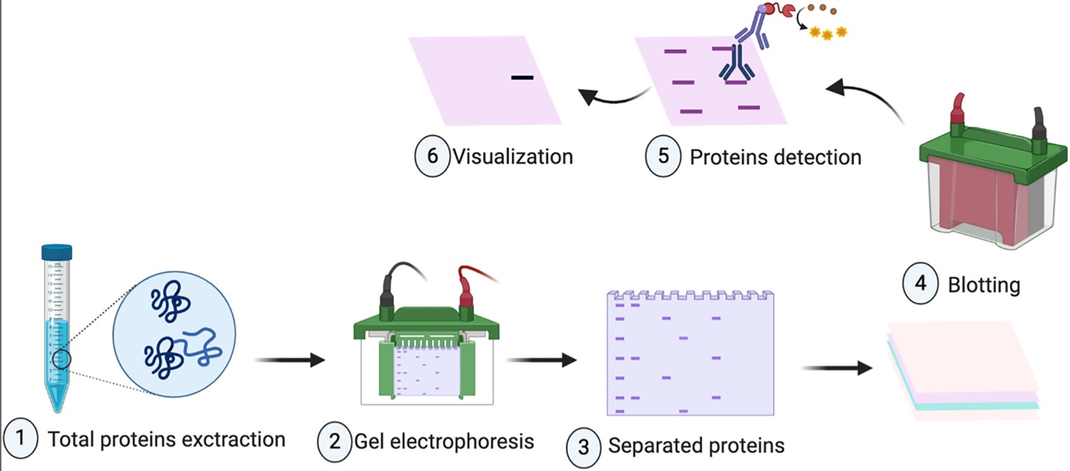
Protein Western blotting is a technique that detects specific proteins in complex samples based on the principle of specific recognition and binding between antigens and antibodies. After extracting total protein from the sample, we use the bicinchoninic acid (BCA) method, the Lowry method, the Bradford method, or the BCA method to determine the total protein concentration. Once quantification is complete, the protein must undergo denaturation. Denaturation refers to the process in which physical factors—such as heating or pressure—and chemical factors—such as strong acids or strong bases—cause changes in the protein’s secondary or tertiary structure, leaving only inactive peptide chains. Typically, in experimental procedures, the secondary and tertiary structures of proteins are denatured by incubating them in a water bath at 96°C or higher for 10 minutes. The proteins in the sample are then separated by molecular weight using polyacrylamide gel electrophoresis (SDS-PAGE). After electrophoresis, the proteins in the gel are transferred to an NC membrane or a PVDF membrane. Why are they transferred to these two types of membranes? This is because NC and PVDF membranes contain microscopic pores invisible to the naked eye. During protein transfer, the proteins enter and bind to these pores. After transfer is complete, a blocking solution is applied to fill the pores that are not occupied by proteins, thereby preventing nonspecific bands, artifacts, and high background during subsequent detection. Since the primary antibody binds to both the target protein and the pores, it is difficult to wash away. it is essential to select a highly specific antibody for the target protein before incubating with the primary antibody. Then, a fluorescently labeled secondary antibody is used to bind to the primary antibody. Finally, a gel imager is used to detect and observe the expression of the target protein in the sample.
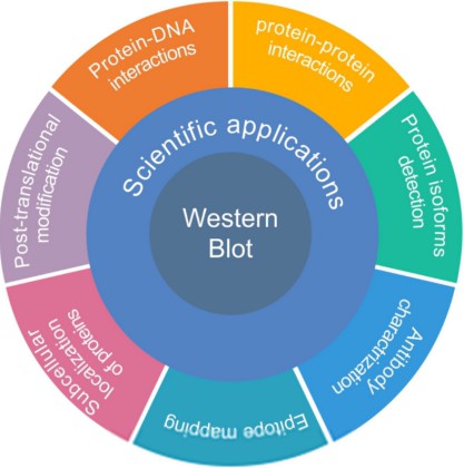
Although the Western blot technique is most commonly used to determine the molecular weight and quantity of proteins in a sample, it also plays an important role in other applications. For example: detecting protein-protein interactions, protein-DNA interactions, and protein-RNA interactions.
KMD Bioscience offers professional protein Western blot (WB) services. Clients need only provide the samples required for the experiment; KMD Bioscience will handle the experimental procedures and data analysis, and ultimately provide a comprehensive experimental analysis report tailored to the client’s project.
- Protein Immunoblotting
- Western Blot
- Protein Interaction Assay
- WB Assay

- Protein Platform
- Protein Expression Services
- Protein Modification & Detection Services
- Protein Interaction Services
- Protein Purification Service
- Antibody Discovery Platform
- VHH Antibody Platform
- Phage Display Platform
- Antibody Humanization Platform
- Single B Cell Screening Platform
- Antibody Expression & Validation Platform
- Peptide Library Platform
- Custom Antibody Platform
- Innovative Drug Discovery Platform
- Product Center
- Proteins
- Antibodies
- Raw Materials Reagents
- Small Molecule Reagents
- Peptide Products
- Nanoparticle Products
- Contact Us
- Phone:400-621-6806
- Mobile:13110936064(WeC
- E-mail:[email protected]
- Address:Room 108, Building 10, No. 6 Hechang Road, Wuqing Development Zone, Tianjin City
-

WeChat
-

Service Account
-
 400-621-6806
 -
 3599163699
 -
 Monday to Friday 9:00-18:00
 -

-
 0
0
-
