


scFv (single-chain variable fragment) has a molecular weight of approximately 25 kDa. It consists of the variable regions of an IgG's light chain (VL) and heavy chain (VH), connected by a flexible linker peptide. scFvs are primarily constructed from hybridoma cells, mouse spleen cells, and human B lymphocytes. They offer advantages including small size, high specificity, and strong affinity. Leveraging their small size, strong tissue penetration, efficient intracorporal clearance, and low immunogenicity, scFvs are widely used for disease diagnosis and therapy. Furthermore, scFvs can be expressed in various systems, such as mammalian cells, yeast cells, plant cells, and insect cells. Among these, the E. coli expression system enables rapid, large-scale scFv production.
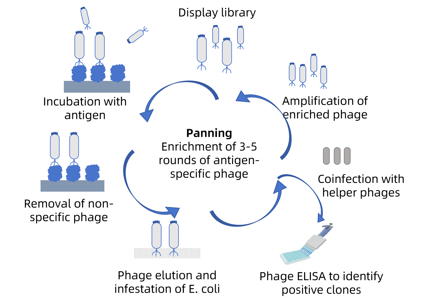
The goal of scFv Antibody Library Screening is to isolate antibody fragments with affinity for the target antigen. Phage Display Library Screening is the most widely applied method for this purpose. The antigen is immobilized on polypropylene microplates. Through 3-5 rounds of panning, scFv-phage antibodies with weak binding are removed, while specific clones binding the antigen are retained. A larger Phage Library Size facilitates the isolation of scFvs with greater diversity and stronger affinity. For Phage Display, the ELISA method is employed to ultimately identify positive clones. For Yeast Display, the FACS method is used, where cells displaying recombinant antibody fragments are detected using labeled antigen.
The goal of scFv Antibody Library Screening is to isolate antibody fragments with affinity for the target antigen. Phage Display Library Screening is the most widely applied method for this purpose. The antigen is immobilized on polypropylene microplates. Through 3-5 rounds of panning, scFv-phage antibodies with weak binding are removed, while specific clones binding the antigen are retained. A larger Phage Library Size facilitates the isolation of scFvs with greater diversity and stronger affinity. For Phage Display, the ELISA method is employed to ultimately identify positive clones. For Yeast Display, the FACS method is used, where cells displaying recombinant antibody fragments are detected using labeled antigen.





 0
0
