


Phage Display Antibody Selection Procedure: An Expert Guide
- 2026-08-16
- 142
Direct Answer: The phage display antibody selection procedure, commonly known as biopanning, is a high-throughput in vitro technique used to isolate high-affinity antibodies from a vast library of billions of variants. The procedure involves four iterative steps: immobilizing the target antigen, incubating it with a library of phage-displayed antibodies to allow binding, washing away non-specific or weak binders, and eluting the strongly bound phages. These eluted phages are then amplified in E. coli to create an enriched pool for the next round of selection. Typically, 3 to 5 rounds are required to isolate highly specific antibody candidates.
Navigating the complex landscape of therapeutic and diagnostic antibody discovery requires moving past outdated, slow methodologies. While traditional hybridoma technology served the industry well for decades, it completely fails when confronted with highly conserved mammalian proteins, toxic antigens, or the urgent need for fully human antibodies. In most professional situations, overcoming these immunological tolerance barriers demands an entirely in vitro approach.
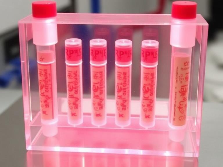
This is where the Phage Display Platform becomes indispensable. However, owning a phage library and knowing how to screen it effectively are two very different realities. The commercial success of a biotherapeutic relies heavily on the stringency and strategic execution of the biopanning process. From our experience at KMD Bioscience, generic protocols yield generic binders with cross-reactivity issues. In this comprehensive guide, we will break down the exact mechanics of the phage display antibody selection procedure, diagnose common screening failures, and provide actionable intelligence on whether you should build these libraries in-house or outsource to specialized experts.
Quick Answer: Key Takeaways
The Core Mechanism: Physical linkage of phenotype (the antibody fragment on the phage surface) and genotype (the DNA inside the phage).
Selection Stringency: Success depends entirely on manipulating the wash conditions. Increasing detergent concentrations and lowering antigen availability across consecutive rounds forces competition for the highest affinity binders.
Library Diversity is King: A selection procedure is only as good as its starting pool. If your initial library diversity is below 10^9 independent clones, your chances of finding a sub-nanomolar binder drop precipitously.
Format Flexibility: The procedure is ideal for generating scFv, Fab, and highly stable nanobodies via a VHH Antibody Platform.
Table of Contents
What is Phage Display Antibody Selection?
Phage display antibody selection is a powerful molecular biology technique that mimics the human immune system in a test tube. Instead of injecting an animal with an antigen and waiting for its B cells to produce antibodies, researchers utilize filamentous bacteriophages (viruses that infect bacteria, typically M13) engineered to display human or animal antibody fragments (like scFv or Fab) on their viral coat proteins.
Because the DNA encoding the specific antibody fragment is encapsulated inside the very same phage that displays it, researchers can physically pull down the binding antibody and immediately sequence its DNA. When you are looking to isolate a therapeutic candidate, this procedure allows you to bypass animal immunization entirely, which is critical when developing fully human antibodies through a direct naive library or an advanced Antibody Humanization Platform.
How the Procedure Works: The Biopanning Workflow
The selection procedure, termed "biopanning," is a cyclical process of enrichment. To isolate a rare, high-affinity antibody from a sea of billions of non-binders, the protocol must be executed with ruthless precision across 3 to 5 rounds. Here is the detailed breakdown of the procedure:

Step 1: Immobilization (Coating). The target antigen is immobilized onto a solid surface, such as an ELISA plate well or magnetic beads. For complex membrane proteins, cells expressing the target antigen can be used directly.
Step 2: Binding (Incubation). The vast phage library (containing up to 10^11 variants) is introduced to the immobilized antigen. Phages with antibody fragments that recognize the antigen will bind to it, while the vast majority remain suspended in solution.
Step 3: Washing (The Critical Differentiator). This is where inexperienced labs fail. The surface is washed extensively with buffers containing detergents (like Tween-20) to remove non-specific and weak binders. From our experience, the stringency of these washes must increase exponentially with each subsequent round to isolate only the tightest binders.
Step 4: Elution. The strongly bound phages are released from the antigen. This is typically achieved by lowering the pH (acid elution) or through competitive elution using an excess of soluble antigen.
Step 5: Amplification. The eluted phages are used to infect E. coli bacteria. The bacteria act as factories, multiplying the successful phages overnight. This newly amplified, enriched pool becomes the starting library for Round 2.
Commercial and Technical Benefits
Is it actually worth investing in a phage display campaign? For modern biopharma, the answer is an uncompromising yes. The primary benefit is absolute control over the selection conditions. Because the selection occurs in vitro, you can manipulate the environment to suit your commercial needs. If you need an antibody that only binds in an acidic tumor microenvironment, you simply perform the washing and binding steps at a low pH.
Furthermore, this procedure allows you to target highly conserved antigens. An animal will not produce antibodies against a protein that is identical to its own (immune tolerance). Phage libraries do not possess an immune system; therefore, they have zero tolerance restrictions, allowing you to generate robust binders against toxic or highly conserved mammalian targets via a Custom Antibody Platform.
Limitations of the Procedure
We must use commercial and practical judgment: phage display is not flawless. The most significant limitation is the loss of natural heavy and light chain pairing. Because the VH and VL genes are randomly recombined during the construction of scFv or Fab libraries, the resulting antibody fragments may exhibit lower stability or solubility than naturally occurring antibodies. Additionally, the procedure is highly susceptible to background noise. If your blocking agent (like BSA or milk) is not thoroughly depleted, you will isolate antibodies against the blocker rather than your target antigen.
Who Should Use It
For commercial users: Pharmaceutical companies developing fully human therapeutic monoclonal antibodies, organizations engineering CAR-T cell therapies requiring stable scFvs, and diagnostic companies looking for ultra-specific nanobodies (VHH) should prioritize phage display. It is the gold standard for high-throughput, customized affinity maturation.
For heavy-duty applications: When working with difficult targets like GPCRs (G protein-coupled receptors) or ion channels, cell-based biopanning via phage display is often the only viable path to discovery.
Who Does Not Need It
For beginners or basic research: If you simply need a basic reagent antibody for a routine Western blot or immunohistochemistry assay against a highly immunogenic, non-toxic protein, a standard polyclonal or hybridoma-derived monoclonal antibody is faster and cheaper. You do not need the immense infrastructure of a phage display platform for standard laboratory reagents.
Common Mistakes in Antibody Selection
In our testing and client rescue projects, we consistently see two fatal errors in the phage display antibody selection procedure. The first is omitting the "subtractive panning" or "depletion" step. Before introducing the library to your target antigen, it must be incubated with the bare solid support and blocking proteins to remove non-specific sticky phages. Failing to do this results in a false-positive final pool.
The second mistake is over-coating the antigen in later rounds. In Round 1, you want a high concentration of antigen to capture any possible binder. But by Round 3, you must drastically reduce the antigen concentration on the plate. This forces the amplified phages to compete fiercely for limited binding sites, ensuring only those with sub-nanomolar affinity survive the procedure.
Buying Considerations: Outsourcing vs. In-House
If you are deciding whether to build a phage library in-house or outsource to a commercial partner, you must evaluate the hidden costs. Constructing a high-quality naive human library with a diversity exceeding 10^9 independent clones requires massive amounts of RNA, highly efficient electroporation protocols, and months of optimization. A poor library will guarantee a failed selection procedure, regardless of your panning skills.
We recommend outsourcing to established platforms that possess pre-validated, high-capacity libraries. Evaluate partners based on their ability to offer parallel screening strategies (e.g., solid-phase vs. solution-phase panning) and their downstream capabilities, such as integration with an Single B Cell Screening Platform for complementary hit generation.
Expert Recommendation
Why Partner with KMD Bioscience?
KMD Bioscience has extensive experience in Recombinant Antibody development and research, supported by a robust technical system that provides a distinct edge within the industry. We deliver premium scientific services to our clients. We have achieved outstanding results in preparing scFv, Fab, and VHH Antibodies, earning the trust of clients globally.
Utilizing Phage Display Antibody Library Construction followed by multiple rounds of ELISA Screening, we efficiently identify high-affinity antibodies for clients. Furthermore, we conduct diverse antibody screening assays tailored to specific client requirements to generate highly specific and stable antibodies. For specialized discovery projects, explore our Peptide Library Platform to accelerate your therapeutic pipeline.
Essential Data Tables
| Parameter | Standard Protocol Target | Why It Matters |
|---|---|---|
| Library Diversity | > 10^9 (Naive) / 10^7 (Immune) | Determines the statistical probability of finding a high-affinity binder. |
| Panning Rounds | 3 to 5 Rounds | Balances maximum enrichment against the risk of losing rare, slow-growing clones. |
| Wash Stringency | Increasing Tween-20 (0.1% to 0.5%) | Physically disrupts weak, non-specific interactions. |
| Elution Method | Trypsin cleavage or Low pH (2.2) | Ensures maximum recovery of tight binders without destroying phage infectivity. |
| Feature | Phage Display Selection | Hybridoma Technology | Single B Cell Screening |
|---|---|---|---|
| Animal Immunization | Not required (if using Naive libraries) | Strictly Required | Strictly Required |
| Target Limitations | None (Can target toxic/conserved antigens) | Fails on toxic or highly conserved targets | Fails on toxic or highly conserved targets |
| Native Chain Pairing | Lost (Random recombination) | Preserved | Preserved |
| Development Timeline | Fast (4-8 weeks from library to hit) | Slow (4-6 months) | Fast (2-4 weeks post-immunization) |
| Pros | Cons |
|---|---|
| Absolute control over selection conditions (pH, temperature, competitors). | Requires highly specialized molecular biology infrastructure and expertise. |
| Ability to generate fully human antibodies directly, skipping humanization steps. | Some eukaryotic proteins may not fold correctly when expressed on bacterial phages. |
| Ideal for selecting highly stable nanobodies (VHH) and scFv fragments. | Risk of isolating fast-growing "parasitic" phages over true high-affinity binders. |
| Evaluation Criteria | What to Demand | Red Flags |
|---|---|---|
| Library Quality Control | Next-Generation Sequencing (NGS) validation of library diversity prior to panning. | Reliance on theoretical diversity calculations without actual sequence validation. |
| Screening Flexibility | Ability to perform both solid-phase (plate) and solution-phase (bead) selection. | Rigid adherence to a single protocol regardless of the antigen's biological nature. |
| Deliverable Transparency | Full sequence data and purified recombinant antibody validation. | Delivering only raw phage lysates without binding kinetics (SPR/BLI) confirmation. |
Frequently Asked Questions
How many rounds of panning are required in a phage display antibody selection procedure?
In most professional situations, 3 to 5 rounds of biopanning are required. Fewer than 3 rounds usually results in a pool overwhelmed by non-specific background binders. More than 5 rounds often leads to a severe loss of diversity, where a single, fast-growing (but not necessarily highest-affinity) phage clone dominates the entire output.
Can phage display be used to find antibodies against membrane proteins?
Yes. While purified soluble proteins are easier to work with, complex membrane proteins (like GPCRs) that lose their conformation when purified can be targeted using cell-based panning. The phage library is incubated directly with cells overexpressing the target membrane protein, accompanied by strict negative depletion against wild-type cells.
Why do we use scFv or Fab formats instead of full IgGs on phages?
Full IgG molecules are too large and complex to be efficiently expressed and assembled in the periplasm of E. coli, which is required for phage assembly. Therefore, smaller fragments like scFv (single-chain variable fragment) or Fab (fragment antigen-binding) are used during the selection procedure. Once the ideal binder is identified, its sequence is easily reformatted into a full IgG for mammalian expression.
Authoritative References & Industry Standards
To ensure alignment with global biopharmaceutical discovery standards, the methodologies discussed in this guide reflect the protocols established and recognized by the following authoritative entities:
National Center for Biotechnology Information (NCBI) / PubMed: The primary repository for peer-reviewed molecular biology protocols, including foundational papers on M13 bacteriophage display mechanics and library construction.
The Antibody Society: An international non-profit association representing individuals and organizations involved in antibody research and development, providing industry benchmarks for therapeutic antibody discovery.
U.S. Food and Drug Administration (FDA): Provides regulatory guidance on the structural characterization, humanization requirements, and preclinical evaluation of monoclonal antibodies derived from in vitro selection platforms.

- Protein Platform
- Protein Expression Services
- Protein Modification & Detection Services
- Protein Interaction Services
- Protein Purification Service
- Antibody Discovery Platform
- VHH Antibody Platform
- Phage Display Platform
- Antibody Humanization Platform
- Single B Cell Screening Platform
- Antibody Expression & Validation Platform
- Peptide Library Platform
- Custom Antibody Platform
- Innovative Drug Discovery Platform
- Product Center
- Proteins
- Antibodies
- Raw Materials Reagents
- Small Molecule Reagents
- Peptide Products
- Nanoparticle Products
- Contact Us
- Phone:400-621-6806
- Mobile:13110936064(WeC
- E-mail:[email protected]
- Address:Room 108, Building 10, No. 6 Hechang Road, Wuqing Development Zone, Tianjin City
-

WeChat
-

Service Account
-
 400-621-6806
-
 3599163699
-
 Monday to Friday 9:00-18:00
-

-
 0
-
