


Phage Display vs Yeast Display: The Definitive Industry Guide for Antibody Discovery
- 2026-07-27
- 173
Selecting the optimal in vitro screening platform is one of the most critical decisions a biotechnology firm must make during early-stage therapeutic development. The debate surrounding phage display vs yeast display is continuous, with commercial viability, development timelines, and target complexity hanging in the balance. In vitro display technologies like phage and yeast display offer a degree of control over the selected antibodies that traditional in vivo methods lack, such as recognizing predefined conformations.
From our experience, many biotech startups burn through crucial funding by utilizing the wrong display system for their specific goals. The failure to align platform strengths with project demands results in lost binders, aggregation issues, and stalled pipelines. In this comprehensive guide, we will break down exactly what these systems are, whether it is actually worth upgrading from one to the other, and how to utilize them for maximum commercial efficacy.
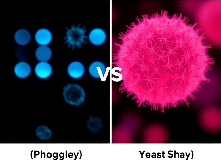
Quick Answer: Which Platform Should You Choose?
If you are deciding between phage display vs yeast display, your choice depends entirely on the stage of your discovery pipeline and your library size requirements.
Choose Phage Display: For primary, naive library screening, nanobody discovery, and peptide isolation. It provides unmatched library diversity (up to 1011 variants) at a significantly lower cost.
Choose Yeast Display: For affinity maturation, fine-tuning lead candidates, and displaying complex eukaryotic proteins that require post-translational modifications (PTMs). It offers precise, quantitative single-cell sorting.
The Verdict: Do not treat them as mutually exclusive. In most professional situations, we recommend a hybrid pipeline: utilize phage display for vast primary discovery, followed by yeast display to mature and optimize the best hits.
Table of Contents
What It Is: Core Technologies Explained
At their core, both platforms genetically fuse a library of proteins to a surface molecule of a host organism, directly linking the genotype (the DNA sequence inside) to the phenotype (the protein displayed outside). However, the biological hosts they employ dictate their capabilities entirely.
Phage Display
Phage display relies on bacterial viruses, predominantly the filamentous bacteriophage M13. Phage display libraries can be constructed by inserting DNA sequences into phage vectors to present the encoded peptide or antibody fragment on the phage surface. Filamentous phages, such as M13, are widely used because they infect Escherichia coli and secrete particles without causing cell lysis, yielding titers up to 1013 per ml. The target protein—often a single-chain variable fragment (scFv), Fab, or nanobody (VHH)—is typically fused to the pIII or pVIII coat proteins. Because it utilizes a rapid prokaryotic system (E. coli), researchers can routinely build libraries containing over 1011 unique variants.
Yeast Display
Yeast display, conversely, utilizes the eukaryotic organism Saccharomyces cerevisiae. The library proteins are fused to the Aga2p cell wall protein, which is tethered to the Aga1p protein anchored in the yeast cell wall. Because yeast is eukaryotic, it possesses complex folding machinery, chaperones, and quality control mechanisms within its endoplasmic reticulum. If a protein misfolds, the yeast cell typically degrades it rather than displaying it. This ensures that the antibodies presented on the surface are structurally sound, though library construction is limited by yeast transformation efficiency, generally capping at 108 to 109 variants.
How It Works: Biopanning vs. FACS Sorting
The mechanics of how these platforms isolate target-binding molecules highlight the most profound differences in the phage display vs yeast display debate.
Phage Selection (Biopanning)
Phage library selection involves iterative biopanning—often on solid phases or magnetic beads—to enrich for binders and reduce non-specific background.
Millions of phages are washed over the immobilized target. Non-binders are washed away, and binders are eluted via pH changes or competitive cleavage.
The eluted phages reinfect E. coli for amplification, and the cycle repeats. While highly effective for pulling rare binders from a massive pool, biopanning is essentially a "blind" process. You cannot easily distinguish between a phage that binds strongly versus one that is simply present in large quantities.
Yeast Selection (FACS)
Yeast display utilizes Fluorescence-Activated Cell Sorting (FACS). The target antigen is fluorescently labeled and incubated with the yeast library in a liquid suspension.
Yeast display offers the distinct advantage of quantitative control during FACS screening, where signal normalization and affinity parameters are precisely defined by flow cytometry boundaries.
Researchers dual-label the cells: one fluorophore measures how much antibody is displayed on the cell surface, and another measures how much antigen is bound. This allows scientists to normalize the binding signal against expression levels, isolating genuinely high-affinity binders in real-time.
Quick Summary Table
| Feature | Phage Display | Yeast Display |
|---|---|---|
| Host Organism | E. coli (Prokaryote) | S. cerevisiae (Eukaryote) |
| Library Size | Massive (1010 - 1011) | Moderate (107 - 109) |
| Screening Method | Biopanning (Solid phase/Beads) | FACS (Flow Cytometry) |
| Primary Strength | De novo discovery, huge diversity | Affinity maturation, complex folding |
The Unique Benefits of Each Platform
When analyzing phage display vs yeast display, you must match the platform's benefits to your specific commercial endgame.
Benefits of Phage Display: The sheer statistical power of phage display cannot be overstated. If you are starting from a naive library without prior immunization, you need raw numbers to find a functional binder. A phage library offers 100 to 1000 times more diversity than a yeast library. Furthermore, a dedicated Phage Display Platform allows for rapid cycle times. A complete screening campaign can be executed in a matter of weeks at a fraction of the cost of eukaryotic systems.
Benefits of Yeast Display: Yeast display excels where phage fails: quantitative precision. In our testing, the ability to sort cells based on real-time kinetic off-rates via FACS allows for affinity maturation down to the picomolar range. Because yeast is eukaryotic, it correctly folds complex human proteins and performs basic post-translational modifications, heavily reducing the risk of aggregation when the antibody is later scaled up in mammalian CHO cells.
Limitations and Structural Bottlenecks
Commercial users must be aware of the inherent bottlenecks before committing funding to a development cycle.
Phage Limitations: Bacteria do not possess mammalian folding chaperones. Many complex human antibodies forced into a phage system will form insoluble inclusion bodies or display with altered conformations. When these hits are reformatted into full IgG molecules for therapeutic use, they frequently lose their binding affinity entirely. Additionally, phage biopanning is susceptible to target-independent "sticky" binders dominating the pool.
Yeast Limitations: Transformation efficiency is the absolute ceiling for yeast display. Generating a library larger than 109 variants requires massive volumes of electroporation, making it prohibitively expensive and time-consuming for initial de novo discovery. Furthermore, yeast hyper-mannosylation can sometimes obscure binding epitopes, creating false negatives during the FACS sorting process.
Who Should Use It & Who Does Not Need It
Who Should Use Phage Display: For heavy-duty applications requiring vast starting diversity, phage display is mandatory. Startups looking for primary hit generation, teams developing robust nanobodies, or researchers isolating synthetic peptides should utilize a specialized Peptide Library Platform based on phage technology.
Who Should Use Yeast Display: Commercial users focused on affinity maturation. If you already have a nanomolar-affinity antibody and need to push it to picomolar affinity for a therapeutic indication, yeast display combined with error-prone PCR is the industry standard.
Who Does Not Need Either: If you are simply looking for a reagent-grade antibody for basic Western blotting or ELISA, building in vitro display libraries is overkill. Traditional immunization and hybridoma techniques remain sufficient. Alternatively, for capturing naturally paired heavy and light chains directly from immunized animals without in vitro bias, leveraging a Single B Cell Screening Platform is far more efficient.
Common Mistakes in Platform Selection
In most professional situations, we see teams make two distinct errors when evaluating phage display vs yeast display:
1. Using Yeast for Naive Discovery: Attempting to build a naive, unimmunized library in yeast often fails because a 108 library simply lacks the statistical diversity to contain high-affinity binders for difficult targets. You will spend months sorting for low-affinity, cross-reactive junk.
2. Relying on Phage for Final Maturation: Once you have a lead candidate, running it through phage display for fine affinity maturation is a mistake. Phage biopanning cannot easily discriminate between a binder with a 5nM Kd and a 1nM Kd. You need the quantitative flow cytometry of yeast display to gate and collect only the slowest-dissociating clones.
Buying Considerations and Commercial Value
When seeking a custom antibody development service, is it worth upgrading to a dual-platform approach? Yes. Outsourcing your discovery requires evaluating the vendor's infrastructure. Ensure the partner you select has proven experience transitioning clones from prokaryotic to eukaryotic systems. If you are developing therapeutics, passing your final yeast or phage hits through an Antibody Humanization Platform is a critical buying consideration to reduce patient immunogenicity.
Pros and Cons Comparison Table
| Platform | Pros (Advantages) | Cons (Disadvantages) |
|---|---|---|
| Phage Display |
|
|
| Yeast Display |
|
|
Platform Buying Guide Matrix
| Project Goal | Target Complexity | Recommended Platform | Why? |
|---|---|---|---|
| De novo discovery (Naive) | Low to Medium | Phage Display | Requires massive diversity to find rare initial binders. |
| Affinity Maturation | High | Yeast Display | Requires FACS to isolate picomolar off-rate improvements. |
| Alpaca VHH Discovery | Medium | Phage Display | Nanobodies fold easily in bacteria without complex PTMs. |
| Complex Human Glycoproteins | Very High | Yeast Display | Requires eukaryotic chaperones to fold correctly on the surface. |
Detailed Technical Comparison Table
| Technical Parameter | Phage Display (M13) | Yeast Display (S. cerevisiae) |
|---|---|---|
| Valency | Monovalent (pIII) or Multivalent (pVIII) | Multivalent (10,000 - 100,000 copies per cell) |
| Discrimination Resolution | Low (Enrichment-based) | High (Single-cell fluorescence resolution) |
| Assay Format Compatibility | Solid phase, Magnetic beads, Cell-panning | Flow Cytometry (FACS), Magnetic sorting (MACS) |
| Typical Turnaround Time | 4 to 6 Weeks | 8 to 12 Weeks |
Expert Recommendation & Integrated Solutions
We recommend abandoning the "either/or" mentality. The most robust therapeutic discovery pipelines employ both platforms sequentially. Use phage display to cast the widest possible net over a massive library of 1011 variants to pull out functional binders. Then, reformat those initial hits, introduce targeted mutagenesis, and move them into a yeast display system for precise FACS-based affinity maturation.
KMD Bioscience possesses an advanced phage display technology platform, offering customized high-quality phage display library construction services. We can create diverse natural libraries (mouse, rabbit, human, etc.), immune libraries (such as VHH libraries, scFv libraries, Fab libraries), synthetic libraries (e.g., peptide libraries), and semi-synthetic libraries for our clients, along with professional library panning and screening services. Through this platform, clients can rapidly identify antibodies or ligands with high affinity and specificity for their target of interest.
Whether you require a highly specialized nanobody library construction service or need to validate your final lead candidates via our Antibody Expression & Validation Platform, choosing a partner with versatile technical infrastructure is paramount. For further updates on how these platforms are evolving in the biopharma sector, we recommend exploring the latest protein research insights.
Frequently Asked Questions
Is phage display or yeast display better for nanobody (VHH) discovery?
Phage display is generally the preferred choice for initial nanobody (VHH) discovery. Because VHH molecules are small, single-domain proteins, they fold exceptionally well in prokaryotic systems without requiring complex mammalian post-translational modifications. Phage display allows you to screen vastly larger immune or naive alpaca libraries much faster than yeast.
Why is yeast display limited in initial library size compared to phage display?
The bottleneck lies in transformation efficiency. Introducing foreign plasmid DNA into eukaryotic yeast cells requires electroporation, and the biological limits of yeast cell walls mean only a fraction of cells successfully take up the DNA compared to highly efficient bacterial (E. coli) transformations used in phage display.
Can yeast display accurately predict expression yields in mammalian systems?
Yes, heavily so. Because yeast uses eukaryotic folding and secretory pathways (including ER quality control), a protein that expresses well and remains stable on a yeast surface has a remarkably high correlation with excellent expression yields when later reformatted into a full IgG and produced in mammalian CHO or HEK293 cells.
How does biopanning differ from FACS sorting?
Biopanning is a bulk physical separation method where millions of phages are washed over an immobilized target, keeping whatever sticks. FACS (used in yeast display) is an analytical single-cell sorting method that uses lasers to evaluate each individual yeast cell for both binding strength and expression levels simultaneously, allowing for precise quantitative isolation.
References & Authoritative Sources
National Center for Biotechnology Information (NCBI) / NIH: Beyond natural antibodies: the power of in vitro display technologies
American Society for Microbiology (ASM) Journals: Phage and Yeast Display
American Chemical Society (ACS) Biomaterials Science & Engineering: Empowering Healthcare with Phage Display Technology

- Protein Platform
- Protein Expression Services
- Protein Modification & Detection Services
- Protein Interaction Services
- Protein Purification Service
- Antibody Discovery Platform
- VHH Antibody Platform
- Phage Display Platform
- Antibody Humanization Platform
- Single B Cell Screening Platform
- Antibody Expression & Validation Platform
- Peptide Library Platform
- Custom Antibody Platform
- Innovative Drug Discovery Platform
- Product Center
- Proteins
- Antibodies
- Raw Materials Reagents
- Small Molecule Reagents
- Peptide Products
- Nanoparticle Products
- Contact Us
- Phone:400-621-6806
- Mobile:13110936064(WeC
- E-mail:[email protected]
- Address:Room 108, Building 10, No. 6 Hechang Road, Wuqing Development Zone, Tianjin City
-

WeChat
-

Service Account
-
 400-621-6806
 -
 3599163699
 -
 Monday to Friday 9:00-18:00
 -

-
 0
0
-
