


How Does Phage Display Differ from Yeast or Other Display Systems?
- 2026-05-27
- 281
Author: KMD Bioscience
Phage display technology remains one of the most powerful tools in modern antibody discovery, protein engineering, and molecular interaction research. However, many researchers entering biologics development often ask an important technical question: how does phage display differ from yeast or other display systems?
From our experience at KMD Bioscience, selecting the correct display platform can significantly impact antibody affinity, screening efficiency, epitope diversity, downstream manufacturability, and overall project success.
While phage display continues dominating large-scale antibody library screening, yeast display, ribosome display, bacterial display, and mammalian display systems each provide unique technical advantages depending on the application.
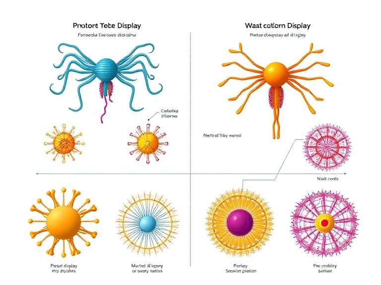
This guide explains how phage display differ from yeast and other display technologies, including workflow differences, screening capabilities, advantages, limitations, and practical CRO considerations for therapeutic antibody discovery.
Table of Contents
Overview of Display Technologies
Display technologies are molecular engineering platforms used to link genotype and phenotype during protein or antibody screening. These systems allow researchers to identify high-affinity binders against specific targets.
The most widely used systems include:
Phage display
Yeast display
Ribosome display
Bacterial display
Mammalian cell display
mRNA display
From our experience, researchers frequently underestimate how much display platform selection affects downstream therapeutic development. The wrong platform can introduce expression challenges, folding artifacts, or affinity limitations later in the discovery pipeline.
KMD Bioscience provides integrated discovery workflows through its Phage Display Platform, helping research organizations accelerate antibody identification and optimization with high-throughput screening strategies.
What Is Phage Display?
Phage display is a molecular display technology where peptides, antibody fragments, or proteins are genetically fused onto bacteriophage coat proteins.
The displayed molecule becomes physically linked to the encoding DNA inside the phage particle, enabling rapid selection of target-binding clones through iterative biopanning cycles.
Core Characteristics of Phage Display
| Feature | Phage Display |
|---|---|
| Host System | Bacteriophage |
| Library Size | Extremely large |
| Screening Speed | Very high |
| Typical Applications | Antibody discovery and peptide screening |
| Common Phage Types | M13, T7, fd |
From our experience, phage display remains the gold standard for large combinatorial antibody library screening because it supports massive diversity and highly efficient enrichment workflows.
Researchers working on recombinant protein optimization often combine display technologies with advanced Protein Expression Platform workflows to improve candidate validation and manufacturability.
What Is Yeast Display?
Yeast display uses genetically engineered yeast cells to present proteins or antibody fragments on the cell surface.
Unlike bacteriophage systems, yeast display offers a eukaryotic folding environment, which improves post-translational processing and conformational accuracy for many proteins.
Core Characteristics of Yeast Display
| Feature | Yeast Display |
|---|---|
| Host System | Saccharomyces cerevisiae |
| Protein Folding | Eukaryotic |
| Flow Cytometry Compatibility | Excellent |
| Library Size | Moderate |
| Affinity Maturation | Highly effective |
From our experience, yeast display performs exceptionally well for affinity maturation because fluorescence-activated cell sorting enables highly precise selection of improved binders.
How Phage Display Differ from Yeast
The most important question researchers ask is how phage display differ from yeast in practical therapeutic discovery.
1. Library Size
Phage display libraries are substantially larger than yeast display libraries.
| Platform | Typical Library Diversity |
|---|---|
| Phage Display | 109 to 1011 |
| Yeast Display | 106 to 108 |
From our experience, larger library diversity gives phage display a major advantage during early-stage antibody discovery against difficult targets.
2. Protein Folding Environment
Yeast display provides a eukaryotic expression environment, enabling improved protein folding and post-translational modifications.
Phage display uses bacterial systems, which may limit folding accuracy for some complex proteins.
This is one of the most important ways phage display differ from yeast display systems.
3. Screening Methodology
Phage display primarily uses biopanning enrichment workflows, while yeast display frequently uses fluorescence-based sorting.
Flow cytometry enables quantitative affinity discrimination in yeast systems.
From our experience, yeast display offers superior precision during affinity maturation stages.
4. Throughput
Phage display supports extremely high-throughput screening with relatively low operational costs.
Yeast display workflows are often more technically demanding and instrument-dependent.
5. Stability and Scalability
Phage particles are highly stable and scalable for industrial library construction.
This scalability remains one of the primary reasons pharmaceutical companies continue relying heavily on phage display.
Other Display Systems Explained
Ribosome Display
Ribosome display is a cell-free system that links proteins to ribosome-mRNA complexes.
Advantages include:
Massive theoretical diversity
No transformation efficiency limitation
Rapid in vitro evolution
However, ribosome complexes are less stable than phage systems.
Mammalian Display
Mammalian display systems provide the closest approximation to native therapeutic antibody expression.
From our experience, mammalian systems are valuable for final-stage validation but are less efficient for ultra-large initial screening.
Projects involving complex protein interactions often integrate advanced Protein Interaction Services to confirm binding specificity after display screening.
Bacterial Display
Bacterial display systems are cost-effective and fast but typically offer lower folding fidelity for complex proteins.
mRNA Display
mRNA display provides extremely large library diversity but requires technically sophisticated workflows and careful RNA stability management.
Advantages and Limitations Comparison
| Display System | Main Advantage | Main Limitation |
|---|---|---|
| Phage Display | Huge library diversity | Bacterial folding limitations |
| Yeast Display | Eukaryotic folding and FACS sorting | Smaller library size |
| Ribosome Display | Cell-free ultra-large diversity | Complex workflow stability |
| Mammalian Display | Native expression environment | High cost and lower throughput |
| Bacterial Display | Fast and inexpensive | Limited folding accuracy |
From our experience, there is no universally superior display technology. The correct system depends on therapeutic target complexity, screening goals, and downstream development priorities.
Best Applications for Each System
When to Use Phage Display
Large antibody discovery campaigns
Peptide library screening
Early-stage binder identification
Industrial-scale screening projects
When to Use Yeast Display
Affinity maturation
Conformational epitope analysis
Eukaryotic protein folding studies
Flow cytometry-based optimization
When to Use Mammalian Display
Complex membrane protein targets
Native glycosylation studies
Late-stage therapeutic optimization
Researchers frequently combine display systems rather than relying on only one platform.
At KMD Bioscience, integrated workflows across the Molecular Platform, Detection Platform, and Process Development Platform help clients transition efficiently from discovery into validation and manufacturability assessment.
How to Select the Right CRO Platform
Selecting the correct CRO partner is just as important as selecting the display system itself.
KMD Bioscience has established a strong technical foundation in therapeutic antibody discovery and molecular engineering since 2022. The company operates ISO9001:2015-certified laboratories and has secured more than 30 patents while supporting global research institutions and biotechnology organizations.
From our experience, researchers should evaluate several factors when choosing display technology providers:
Library diversity capability
Screening throughput
Protein engineering expertise
Analytical validation systems
Manufacturing scalability
Regulatory documentation quality
Complex biologics projects may also require integration with advanced Gene Editing Platform and Plant Genetic Transformation Platform technologies depending on downstream research goals.
Summary Table
| Feature | Phage Display | Yeast Display | Mammalian Display |
|---|---|---|---|
| Library Size | Very large | Moderate | Smaller |
| Folding Accuracy | Moderate | High | Very high |
| Screening Speed | Very fast | Moderate | Slower |
| Cost | Lower | Moderate | Higher |
| Affinity Maturation | Good | Excellent | Good |
| Industrial Scalability | Excellent | Good | Moderate |
Conclusion
Understanding how phage display differ from yeast and other display systems is essential for successful antibody engineering and biologics discovery.
From our experience at KMD Bioscience, phage display remains unmatched for large-scale screening diversity and industrial scalability, while yeast display excels in affinity maturation and conformational precision.
No single platform solves every discovery challenge. The most successful therapeutic programs frequently combine multiple display technologies to maximize screening efficiency, binding quality, and downstream manufacturability.
As antibody engineering continues evolving in 2026, integrated CRO partnerships with broad platform expertise will become increasingly important for accelerating therapeutic innovation.
FAQs
What is the biggest difference between phage display and yeast display?
The biggest difference is that phage display offers much larger library diversity, while yeast display provides better eukaryotic protein folding and flow cytometry-based selection.
Why is phage display widely used in antibody discovery?
Phage display supports massive combinatorial libraries and highly scalable screening workflows, making it ideal for therapeutic antibody discovery.
Is yeast display better for affinity maturation?
Yes. From our experience, yeast display performs exceptionally well during affinity maturation because of precise fluorescence-based sorting.
Can multiple display systems be combined?
Absolutely. Many discovery programs use phage display for initial screening and yeast display for later optimization.
Which display system is closest to native human protein expression?
Mammalian display systems provide the closest approximation to native human protein folding and glycosylation.
References

- Protein Platform
- Protein Expression Services
- Protein Modification & Detection Services
- Protein Interaction Services
- Protein Purification Service
- Antibody Discovery Platform
- VHH Antibody Platform
- Phage Display Platform
- Antibody Humanization Platform
- Single B Cell Screening Platform
- Antibody Expression & Validation Platform
- Peptide Library Platform
- Custom Antibody Platform
- Innovative Drug Discovery Platform
- Product Center
- Proteins
- Antibodies
- Raw Materials Reagents
- Small Molecule Reagents
- Peptide Products
- Nanoparticle Products
- Contact Us
- Phone:400-621-6806
- Mobile:13110936064(WeC
- E-mail:[email protected]
- Address:Room 108, Building 10, No. 6 Hechang Road, Wuqing Development Zone, Tianjin City
-

WeChat
-

Service Account
-
 400-621-6806
 -
 3599163699
 -
 Monday to Friday 9:00-18:00
 -

-
 0
0
-
