


Membrane-Lytic Antimicrobial Peptide PmH2A-AMP (5-13)[KLLK]3: A Promising Molecule for Aquatic Antimicrobial Applications
- 2026-04-02
- 153
Journal: | Fish and Shellfish Immunology |
| Impact Factor: | 4.8 (2023) |
| Client Institution: | Fisheries College, Guangdong Ocean University; Guangdong Provincial Key Laboratory of Aquatic Animal Disease Control and Healthy Aquaculture |
| Authors: | Dr. Chenghao Shen and Dr. Zhijie Guo from Guangdong Ocean University are the co-first authors of the paper. Professor Haiying Liang is the corresponding author. Guangdong Ocean University is the primary institution. |
Antimicrobial peptides (AMPs) are key components of the innate immune system and serve as core defense molecules that protect aquatic animals from pathogenic microorganism infections. The pearl oyster Pinctada fucata martensii is an economically important bivalve species in marine pearl culture. Research on histone-derived antimicrobial peptides within its innate immune system provides both a crucial theoretical foundation and practical references for disease control in bivalve aquaculture.
In 2023, a research team from Guangdong Ocean University published findings in Fish and Shellfish Immunology that revealed, for the first time, the structural characteristics, antimicrobial activity, and immune response mechanisms of H2A histone-derived antimicrobial peptides (PmH2A‑AMPs) from Pinctada fucata martensii. The researchers synthesized and structurally optimized these peptides to obtain highly active antimicrobial derivatives. They confirmed that these peptides exert their antimicrobial function by disrupting cell membrane structures and are also involved in an immune response related to neutrophil extracellular traps (NETs). This work provides a novel research direction and potential targets for the development and application of antimicrobial peptides in aquaculture.
I. Overview
This study focused on the H2A histone protein (PmH2A) of the pearl oyster Pinctada fucata martensii to address the lack of sequence information and unclear functional mechanisms of antimicrobial peptides in this species. A comprehensive investigation was conducted, encompassing the design and synthesis of antimicrobial peptides, evaluation of in vitro antimicrobial activity, elucidation of antimicrobial mechanisms, and analysis of in vivo immune responses.
Based on the sequence characteristics of Buforin II, the research team designed and synthesized three PmH2A-derived antimicrobial peptides through amino acid substitution, N-terminal truncation, and motif repetition: PmH2A-AMP, PmH2A-AMP (5-21), and PmH2A-AMP (5-13)[KLLK]3. The minimum inhibitory concentrations (MICs) of these three peptides against seven Gram-positive and Gram-negative bacterial strains were determined using the broth microdilution method. Morphological changes in bacteria following peptide treatment were observed via transmission electron microscopy (TEM), confirming that their antimicrobial mechanism involves membrane disruption. Using quantitative real-time PCR (qRT-PCR) and immunohistochemistry (IHC), the spatiotemporal expression patterns of PmH2A in the hemocytes and gill tissues of Pinctada fucata martensii were analyzed following stimulation with three pathogen-associated molecular patterns (PAMPs): lipopolysaccharide (LPS), peptidoglycan (PGN), and polyinosinic-polycytidylic acid (Poly (I:C)). The results demonstrated that LPS induces the release of antimicrobial histones in Pinctada fucata martensii, triggering a NETs-like immune response.
II. Mechanism of Membrane Disruption by Antimicrobial Peptides
Observation via transmission electron microscopy (TEM) revealed morphological changes in bacteria following peptide treatment. In the PBS control group, Vibrio parahaemolyticus cells remained intact with uniformly distributed cytoplasm. In contrast, treatment with PmH2A-AMP resulted in no significant alterations to the cell wall or overall cellular morphology; however, damage to the inner membrane was evident, characterized by slight invagination and uneven cytoplasmic distribution. These findings confirm that the antimicrobial action of PmH2A-AMP targets the bacterial inner membrane, disrupting its integrity and altering membrane permeability, thereby inhibiting bacterial growth.
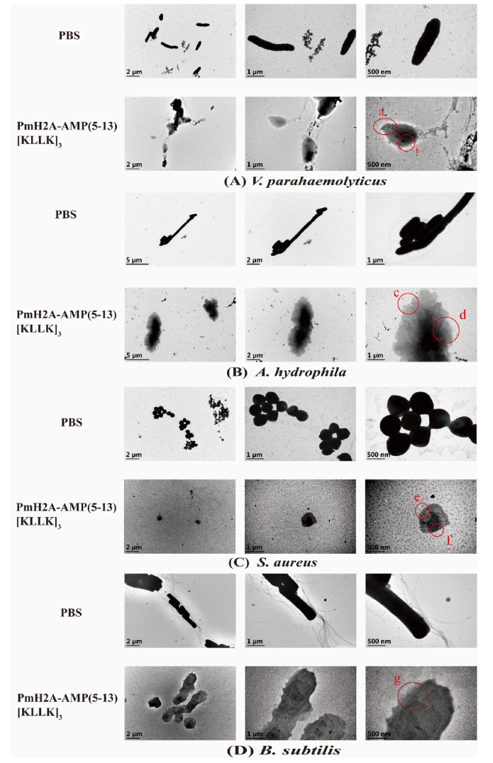
Fig 1. Morphological changes in different bacterial species upon exposure to PmH2A- AMP
III. [KLLK]3 Cationic Repeat Motif Enhances Membrane Disruption Efficiency
TEM results demonstrated that PmH2A-AMP (5-13)[KLLK]³ induced significant morphological damage in Vibrio parahaemolyticus, Aeromonas hydrophila, Staphylococcus aureus, and Bacillus subtilis, with distinct differences in the damage patterns observed between Gram-negative and Gram-positive bacteria. This peptide exhibits broad-spectrum membrane-disruptive activity. The incorporation of the [KLLK]³ repeat motif significantly enhanced its capacity to disrupt bacterial membrane structures, representing a key factor underlying its substantially higher antimicrobial activity compared to the original peptide.
![Fig 2. Morphological changes in different bacterial species upon exposure to PmH2A-AMP(5–13)[KLLK]3.png](/uploads/images/20260402/1775123985143201.png)
Fig 2. Morphological changes in different bacterial species upon exposure to PmH2A-AMP(5–13)[KLLK]3
IV. mRNA Expression Profiles of PmH2A in Hemocytes and Gill Tissues Following PAMP Stimulation
qRT-PCR analysis revealed that PmH2A mRNA expression in the hemocytes and gill tissues of Pinctada fucata martensii exhibited distinct spatiotemporal specificity following stimulation with pathogen-associated molecular patterns (PAMPs). Variations in induction efficacy were observed among different PAMPs, with lipopolysaccharide (LPS) demonstrating the most pronounced inductive effect. Hemocytes function as the initial immune site for PmH2A in response to pathogen invasion, while gill tissues serve as the primary effector organ for its antimicrobial activity. PmH2A is primarily involved in the immune response against bacterial infections and displays expression patterns indicative of a secondary immune response in both hemocytes and gill tissues.
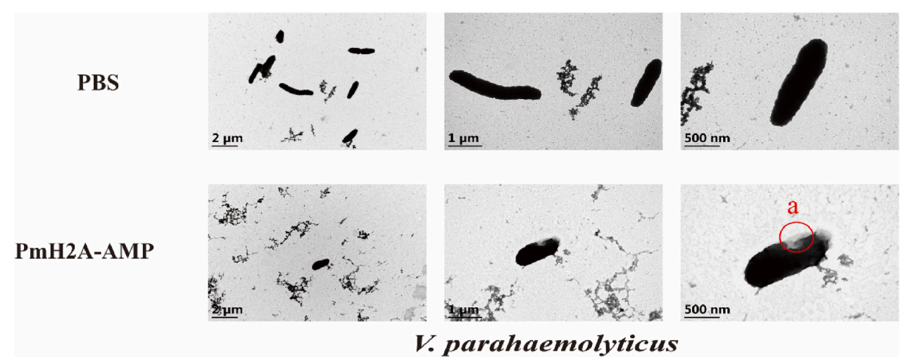
Fig 3. The regulatory role of PmH2A in the blood cells and gills of Martin's flagellate towards pathogen-associated molecular patterns
V. Spatiotemporal Expression Profile of PmH2A-AMP Protein in Gill Tissues Following LPS Stimulation
Immunohistochemical analysis revealed a time-dependent expression pattern of the PmH2A-AMP protein in the gill tissues of Pinctada fucata martensii following LPS stimulation. These findings validate the spatiotemporal expression of PmH2A at the protein level, corroborating the mRNA expression results obtained via qRT-PCR. This confirms that gill tissues serve as the primary effector organ for the antimicrobial activity of PmH2A. The regulation of its protein expression occurs predominantly at the transcriptional level, and the specific high expression observed in gill filament epithelial cells reflects the spatial specificity of immune defense. Furthermore, this provides protein-level evidence supporting the existence of a histone-mediated NETs-like innate immune strategy in Pinctada fucata martensii.
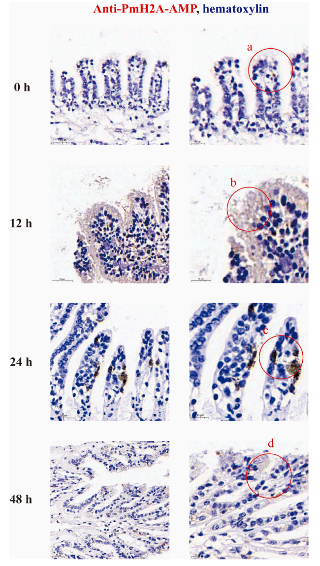
Fig 4. Distribution of PmH2A-AMP in gill tissues after LPS stimulation
This study reports the first identification of the PmH2A gene in Pinctada fucata martensii and the synthesis of three classes of gene-derived antimicrobial peptides. Among these, PmH2A‑AMP (5‑13)[KLLK]³ was identified as a highly effective, broad-spectrum antimicrobial peptide that exerts its bactericidal effects via membrane disruption. PAMPs were shown to induce the sequential expression of PmH2A in hemocytes and gill tissues, forming an immune regulatory pathway characterized by rapid response, circulatory transport, and localized defense. These findings systematically elucidate the molecular mechanisms underlying the innate immunity of Pinctada fucata martensii and provide a natural template for the development of novel antimicrobial agents.
The three peptides used in this study—PmH2A-AMP, PmH2A-AMP (5–21), and PmH2A-AMP (5–13)[KLLK]³—were synthesized at KMD Bioscience (Tianjin) using Fmoc-based solid-phase chemistry. Following synthesis, the target peptides were purified by reversed-phase high-performance liquid chromatography (RP-HPLC) for subsequent use in peptide library screening and related activity studies.
KMD Bioscience possesses an advanced phage display peptide library construction and screening platform capable of generating ultra-high throughput libraries with titers up to 1013 pfu/mL. Leveraging this platform, we offer not only a variety of pre-made peptide libraries (such as linear 7-mer, 12-mer, 15-mer, and cyclic 7-mer libraries) but also custom peptide library construction services tailored to specific research objectives. Our comprehensive workflow encompasses library construction, high-throughput screening, and sequencing validation, supporting your research from start to finish.
- Antimicrobial Peptides
- Membrane Disruption
- Phage Display Library

- Protein Platform
- Protein Expression Services
- Protein Modification & Detection Services
- Protein Interaction Services
- Protein Purification Service
- Antibody Discovery Platform
- VHH Antibody Platform
- Phage Display Platform
- Antibody Humanization Platform
- Single B Cell Screening Platform
- Antibody Expression & Validation Platform
- Peptide Library Platform
- Custom Antibody Platform
- Innovative Drug Discovery Platform
- Product Center
- Proteins
- Antibodies
- Raw Materials Reagents
- Small Molecule Reagents
- Peptide Products
- Nanoparticle Products
- Contact Us
- Phone:400-621-6806
- Mobile:13110936064(WeC
- E-mail:[email protected]
- Address:Room 108, Building 10, No. 6 Hechang Road, Wuqing Development Zone, Tianjin City
-

WeChat
-

Service Account
-
 400-621-6806
 -
 3599163699
 -
 Monday to Friday 9:00-18:00
 -

-
 0
0
-
