


A Guide to High-Throughput Screening of VHH Antibody Libraries: Key Parameter Control That Determines Success or Failure During the Screening Process
- 2026-09-14
- 26
VHH is a single-domain antibody derived from the variable region of heavy-chain antibodies in camelids. It has a molecular weight of approximately 13–15 kDa and consists of three complementarity-determining regions (CDRs), among which the CDR3 loop contributes most significantly to antigen binding[1]. This unique structural feature confers excellent stability to VHH antibodies, enabling them to withstand extreme pH levels and high temperatures during phage library screening, as well as more rigorous washing conditions, making them particularly suitable for the biological selection of whole-cell surface antigens.
In the VHH antibody discovery process, phage library screening is one of the most critical technical platforms. However, many adjustable parameters in the biopanning process are often applied in a “one-size-fits-all” manner, resulting in low screening efficiency. This article breaks down the VHH antibody library screening process into eight quantifiable key parameters and systematically analyzes the optimization strategies for each parameter in phage library screening, helping researchers obtain a higher proportion of functional VHH antibody-positive clones during biopanning.
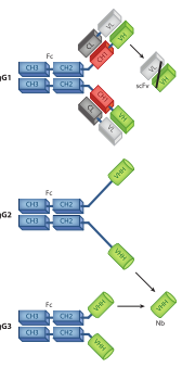
Figure 1: Schematic Diagram of the VHH Structure[1]
I. Antigen coating density
(i) The Nature of the Problem
The coating density of antigens on a solid-phase carrier directly determines the number and types of epitopes accessible to VHH. Excessively high density may lead to epitope masking, while excessively low density may result in the loss of low-abundance, high-affinity clones. Furthermore, due to their small size and unique epitope-binding mode, VHHs have different antigen density requirements than standard antibodies.
(ii) Decision-Making Logic
Table 1: Recommended Antigen Coating Densities
Round | Strategy | Density Recommendations |
Round 1 | High Density | 5-10 μg/mL(or 100–200 pmol/well) |
Subsequent rounds | Decreasing by rounds | Drop to 1–0.2 μg/mL |
II. Negative Selection
(i) The Nature of the Problem
Phage display screening enriches target-binding clones and depletes all other clones by applying selection pressure. However, in practice, phages that bind nonspecifically continue to interfere with the enrichment of the target antibody. The primary purpose of negative screening is to remove phages that bind to non-target molecules prior to positive screening, thereby significantly improving the efficiency and specificity of positive screening.
(ii) Negative Target Selection Strategy
Table 2: Types of Negative Target Selection Strategies
Negative Target Type | Applicable Scenarios | Purpose |
BSA/Skim Milk | Eliminating nonspecific binding of the blocking agent | Basic Purification |
Tag protein | Fusion Protein Antigen | Removal of tag-binding clones |
Homologous proteins | Multimember protein families | Distinguishing Specific Clones |
Antigen-negative cells | Whole-cell screening | Removal of nonspecific binders to membrane proteins |
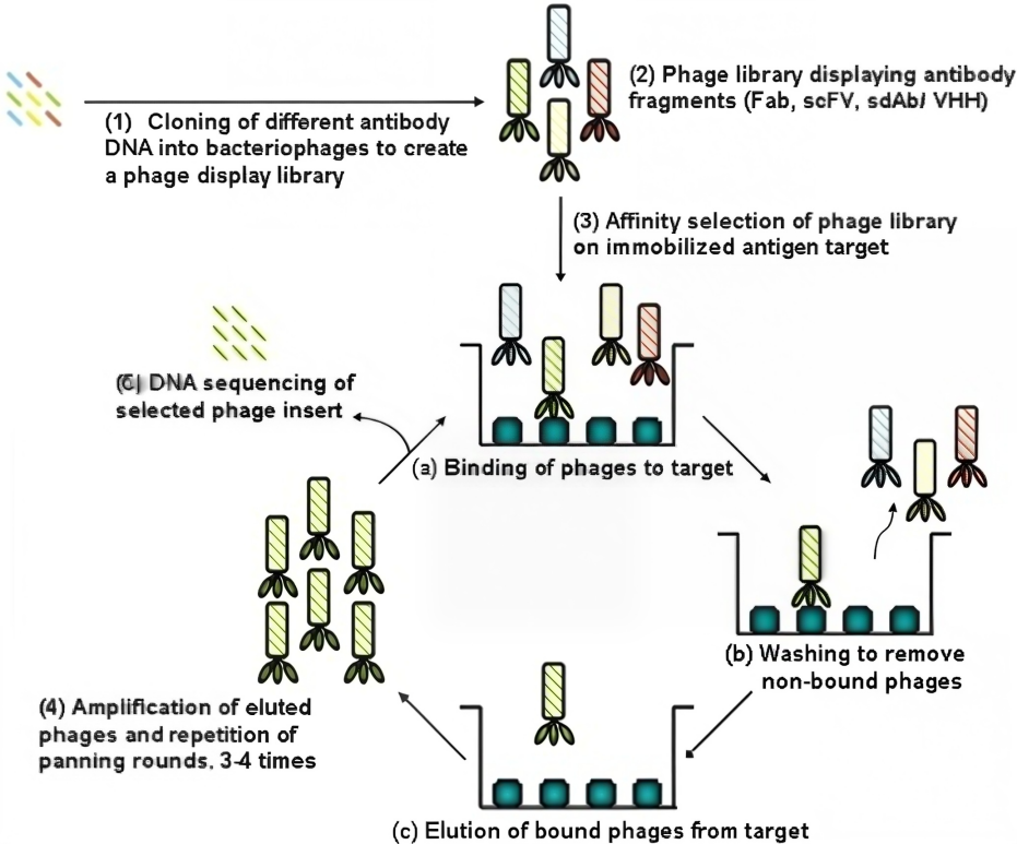
Figure 2: Schematic Diagram of the Phage Display Screening Process[2]
III. Number of Selection Rounds
(i) The Nature of the Problem
The number of rounds in biopanning directly affects the enrichment of VHH antibodies. If the number of rounds is too low, the phage library is not screened thoroughly; if the number of rounds is too high, the diversity of the VHH library drops sharply, and clones with unique binding epitopes may even be lost.
(ii) Optimization Suggestions
Ø Immune library: High-affinity VHHs can typically be obtained after three rounds of screening. In a study on HPV16 E7, high-affinity VHHs were obtained after three rounds of biopanning.
Ø Natural/synthetic libraries: Typically require 3–4 rounds
Ø Whole-cell screening: Some studies have conducted as many as eight rounds of selection
IV. Washing Severity
(i) The Nature of the Problem
Washing is the most critical step in the screening process—it directly determines the extent to which nonspecific phages are removed and the efficiency with which high-affinity clones are retained.
(ii) Optimization Suggestions
Table 3: Washing Control Strategies
Round | Tween-20 concentration | Number of washes | Washing Time |
Round1 | 0.05% | 3-5 | 2min |
Round2 | 0.1% | 5-8 | 5min |
Round3 | 0.1%-0.5%[3] | 8-15 | 5-10min |
V. Antigen Dosage
(i) The Nature of the Problem
In the screening of phage libraries for VHH antibodies, the amount of antigen added directly determines the “selective pressure” of the biological selection process. A high antigen dose preserves VHH diversity but exerts low affinity pressure; a low antigen dose exerts high affinity pressure but may result in the loss of low-abundance VHH antibody clones.
(ii) Optimization Suggestions
Ø First round (to ensure diversity): Add 100–200 pmol of antigen
Ø Final round (affinity binding): Add 10–20 pmol of antigen
It is important to note that, due to their small molecular weight (~15 kDa), VHHs bind to antigens with a different stoichiometry than conventional antibodies. When the antigen has a small molecular weight (e.g., <30 kDa), the amount of antigen used should be appropriately increased to ensure that sufficient epitopes are exposed.
VI. Elution Conditions
(i) The Nature of the Problem
Elution is the most critical step in the bio-screening process—it directly determines the extent to which nonspecific phages are removed during phage library screening, as well as the efficiency with which high-affinity VHH antibody clones are retained. Because VHH antibodies are structurally extremely stable, they can withstand more rigorous washing conditions than scFvs, which is one of their key advantages in phage library screening.
(ii) Optimization Suggestions
Table 4: Recommendations for Elution Method Optimization
Elution Method | Mechanism | Advantages | Disadvantages |
Acid Elution | Low pH disrupts protein-antigen interactions | Highly versatile | May impair some cloning activity |
Trypsin Elution | Cleaving Bacteriophage Proteins | Minimal damage to the VHH structure | Trypsin must be inactivated |
Competitive Elution | Free antigen-binding sites | Enriching True Functional Clones | Requires a large amount of antigen and is a complex procedure |
VII. Selection of Host Bacteria
(i) The Nature of the Problem
During phage library screening, phage particles must be transformed into host bacteria to allow for phage packaging and amplification. Transformation efficiency and phage packaging capacity vary significantly among different host bacteria, which directly affects the representativeness of the VHH library and the recovery rate of products from biological selection[4].
(ii) Optimization Suggestions
Ø TG1: The most widely used VHH phage display host, with high electroporation efficiency
Ø SS320: Suitable for electroporation of large-scale libraries
Ø CM13K trypsin-sensitive auxiliary phage: Can be used in protocols requiring trypsin elution
VIII. Number of copies in the Input library
(i) The Nature of the Problem
In each round of biological selection from a phage library, the number of input phages determines whether the diversity of the VHH library is adequately represented.
(ii) Optimization Suggestions
The number of input phages for each round of biopanning should be at least 10 times the capacity of the VHH antibody library.
High-throughput screening of VHH antibody libraries is, at its core, an art form in which eight parameters work in concert. From antigen coating density, negative selection, the number of screening rounds, wash stringency, antigen loading, elution conditions, host strain selection, to the number of input copies—each parameter warrants careful evaluation before the project begins and optimization round by round throughout the screening process.In this workflow, all eight key parameters should be dynamically adjusted round by round based on NGS feedback, forming a closed-loop control system.
KMD Bioscience possesses the technical platforms and service capabilities to address every key parameter in VHH antibody library screening, enabling us to provide clients with one-stop VHH antibody discovery services ranging from antigen design, animal immunization, library construction and screening, to functional validation. We strictly implement a dynamic optimization strategy: based on target characteristics, library type, and the client’s end-use scenario, we tailor the optimal combination of solutions for each project, ensuring clients do not waste valuable time on trial-and-error parameter adjustments.
FAQs
Q1:Why is the “washing” step so crucial—so much so that it’s even called the most decisive step?
In the “adsorption–washing–elution–amplification” cycle, the goal of the washing step is to remove phages that bind weakly to the target or lack specific binding altogether. By progressively increasing the “intensity” of the washing step—for example, by increasing the number of washes, raising the concentration of surfactants (such as Tween-20) in the buffer, or extending the duration of each wash—the “threshold” for the screening can be gradually raised.
Q2: Since the CDR3 loop of VHH contributes most to antigen binding, are there any specific requirements regarding antigen coating density?
Since VHH relies solely on the CDR3 loop to mediate most antigen contact, its binding “footprint” is much smaller than that of conventional antibodies. The CDR3 loop of VHH can adopt three conformations—Upright, Half-Roll, and Roll—and these different conformations determine how the nanobody interacts with its target. Based on this structural feature, specific requirements for antigen coating density include:
Ø Excessively high antigen coating density can mask the CDR3 binding site. When antigen molecules are densely packed on a solid-phase surface, steric hindrance between adjacent antigens may directly prevent the VHH CDR3 loop from approaching its epitope. Therefore, the antigen coating density for VHH screening should generally be lower than that for scFv or Fab screening.
Ø Conversely, an antigen coating density that is too low may result in the loss of low-abundance clones. The CDR3 loop of VHH is long and flexible, enabling it to access recessed or conformational epitopes that conventional antibodies cannot reach. However, if the antigen density is too low, these highly specific but low-abundance VHH clones may not have the opportunity to come into contact with the antigen at all.
Ø A concentration range of 5–10 μg/mL in the first round, reduced to 1–0.2 μg/mL in subsequent rounds, is generally suitable for soluble protein antigens with molecular weights between 30 and 100 kDa. For small-molecule antigens (<10 kDa, such as haptenes and peptides), due to the limited number of epitopes on a single antigen molecule, a higher coating density (e.g., 10–20 μg/mL) may be required to ensure sufficient exposure of binding sites; for large-molecule antigens (>150 kDa), the density can be appropriately reduced to avoid steric hindrance.
Q3: When “decreasing antigen coating density with each round” and “increasing wash severity with each round” are performed simultaneously, how can you determine which parameter is causing the problem?
These two parameters affect the O/I ratio in different ways; the source of the problem can be identified using the following method:
Ø Monitor the absolute titer of the eluate rather than relying solely on the O/I ratio. If the antigen coating density drops too low, the absolute phage count in the eluate will decrease significantly (below 10⁴–10⁵ CFU); conversely, if the washing is simply too aggressive, the absolute count in the eluate may remain normal, but the O/I ratio will not increase after subsequent amplification.
Ø Establish an “intermediate monitoring indicator”—the proportion of phage remaining after washing. After each wash, take a small sample of the wash buffer to measure the titer and observe the phage elution curve over the course of each wash cycle. If an abnormally high number of phages are eluted in the early wash supernatants (first 3 washes) of a given round, this suggests that antigen coating may be insufficient; if a large number of phages continue to be eluted in the late wash supernatants (subsequent washes), this suggests that the washing may be too aggressive.
Ø Refer to typical data ranges. In the screening of CD19-specific VHHs, the O/I ratio increased from 7×10⁻⁶ in the first round to 1.37×10⁻⁴ in the fifth round, with an enrichment factor of 19.5-fold. If the O/I ratio in a given round does not increase or even decreases compared to the previous round, and the input quantity is normal, priority should be given to reducing the antigen coating density (rather than adjusting the washing intensity), as washing intensity typically only needs to be increased when the O/I ratio fails to improve for two consecutive rounds.
Q4: What are the key differences in parameter control between whole-cell screening and purified protein screening?
Whole-cell screening and purified protein screening exhibit systematic differences in the regulation of eight parameters:
Ø Antigen “coating density” cannot be precisely controlled. The level of antigen expression on the cell surface is determined by the cell line and cannot be adjusted as precisely as with purified proteins. The solution is to select cell lines with different expression levels (high-expression vs. low-expression) or to quantify cell surface antigen density using antibody-labeled flow cytometry.
Ø Negative selection is even more critical. In whole-cell screening, the sources of nonspecific binding encountered by phages are extremely complex (cell membrane components, culture medium components, cell debris, etc.). Therefore, antigen-negative cells must be used as negative selection controls. Some studies have employed a “negative selection → positive selection” serial mode repeated for four rounds.
Ø More selection rounds are required. Whole-cell screening involves higher levels of noise and typically requires more rounds to achieve effective enrichment—some studies have conducted as many as 8 rounds.
Ø Different washing strategies. High-concentration detergents cannot be used in whole-cell screening (as they would compromise cell integrity); the rigor of washing is primarily achieved by increasing the number of washes (rather than raising the Tween-20 concentration).
Ø Different elution methods. Whole-cell screening often employs a two-step elution process using acid and alkali to maximize the recovery of phages bound to different regions of the cell surface.
Q5: How can NGS be used in conjunction with traditional selection-based parameter regulation?
NGS can serve as a “microscope” for traditional parameter-controlled screening—it not only reveals “how much has been enriched” (the O/I ratio) but also shows “what has been enriched” (dynamics at the sequence level).
Specific ways in which NGS data guides parameter adjustments:
Ø Monitor the rate of diversity loss. If NGS data show that the number of unique CDR3 sequences drops sharply from 10⁴ to 10² after a given round of selection (indicating a steep decline in diversity), this suggests that the selection pressure in the current round (possibly due to excessively harsh washing conditions or insufficient antigen input) is too severe. In the next round, the washing conditions should be relaxed or the antigen input increased to “rescue” the diversity.
Ø Identify early enrichment signals. NGS can detect an increase in the frequency of certain sequence families as early as after the first round of selection—these “early enrichment signals” can guide parameter adjustments for the second round. If a particular family is already significantly enriched in the first round, the stringency can be appropriately increased in the second round to accelerate its purification.
Ø Distinguish between “true positives” and “amplification bias.” Certain sequences may be enriched due to high PCR amplification efficiency rather than strong binding affinity—NGS can detect this “amplification bias.” If the abundance of a sequence across selection rounds does not align with trends in the O/I ratio, be wary of the possibility of amplification bias.
Ø Determine when to terminate the selection process. When NGS analysis shows that, after a given round of selection, the top 10 sequence families account for more than 80% of the total sequences, and the sequence composition in subsequent rounds no longer changes significantly, this indicates that the selection has reached convergence and can be terminated.
NGS-Assisted Screening Workflow:
Round 1 screening → Collect output samples for NGS (while proceeding with Round 2) → NGS data analysis (approximately 5–7 days) → Adjust parameters for Round 3 based on NGS results → Round 3 screening → Re-verify via NGS → Screening concludes.
[1]Muyldermans S. Nanobodies: natural single-domain antibodies. Annu Rev Biochem. 2013;82:775-97. doi: 10.1146/annurev-biochem-063011-092449. Epub 2013 Mar 13. PMID: 23495938.
[2]Yun YH, Eteshola E, Bhattacharya A, Dong Z, Shim JS, Conforti L, Kim D, Schulz MJ, Ahn CH, Watts N. Tiny medicine: nanomaterial-based biosensors. Sensors (Basel). 2009;9(11):9275-99. doi: 10.3390/s91109275. Epub 2009 Nov 19. PMID: 22291565; PMCID: PMC3260642.
[3]Qian M, Xiao S, Yang Y, Yu F, Wen J, Lu L, Wang H. Screening and identification of cyprinid herpesvirus 2 (CyHV-2) ORF55-interacting proteins by phage display. Virol J. 2023 Apr 12;20(1):66. doi: 10.1186/s12985-023-02026-x. PMID: 37046316; PMCID: PMC10091560.
[4]Eyssen, L. E. A., Ramadurai, S., Abdelkarim, S., Buckle, I., Cornish, K., Lin, H., Jones, A. K., Stephens, G. J. and Owens, R. J. (2024). From Llama to Nanobody: A Streamlined Workflow for the Generation of Functionalised VHHs. Bio-protocol 14(6): e4962. DOI: 10.21769/BioProtoc.4962.
- VHH nanobody
- Phage display biopanning
- Eight key parameters

- Protein Platform
- Protein Expression Services
- Protein Modification & Detection Services
- Protein Interaction Services
- Protein Purification Service
- Antibody Discovery Platform
- VHH Antibody Platform
- Phage Display Platform
- Antibody Humanization Platform
- Single B Cell Screening Platform
- Antibody Expression & Validation Platform
- Peptide Library Platform
- Custom Antibody Platform
- Innovative Drug Discovery Platform
- Product Center
- Proteins
- Antibodies
- Raw Materials Reagents
- Small Molecule Reagents
- Peptide Products
- Nanoparticle Products
- Contact Us
- Phone:400-621-6806
- Mobile:13110936064(WeC
- E-mail:[email protected]
- Address:Room 108, Building 10, No. 6 Hechang Road, Wuqing Development Zone, Tianjin City
-

WeChat
-

Service Account
-
 400-621-6806
 -
 3599163699
 -
 Monday to Friday 9:00-18:00
 -

-
 0
0
-
